{"title":"Neuro-Ophthalmological Optic Nerve Cupping: An Overview.","authors":"Ethan Waisberg, Jonathan A Micieli","doi":"10.2147/EB.S272343","DOIUrl":null,"url":null,"abstract":"<p><p>Optic nerve cupping or enlargement of the cup-to-disc ratio is widely recognized as a feature of glaucoma, however it may also occur in non-glaucomatous optic neuropathies. The most well-recognized non-glaucomatous optic neuropathies that cause cupping include compressive optic neuropathies, arteritic anterior ischemic optic neuropathies, hereditary optic neuropathies, and optic neuritis. Cupping is thought to consist of two main components: prelaminar and laminar thinning. The former is a shallow form of cupping and related to loss of retinal ganglion cells, whereas the latter involves damage to the lamina cribrosa and peripapillary scleral connective tissue. Differentiating glaucomatous and non-glaucomatous optic nerve cupping remains challenging even for experienced observers. Classically, the optic nerve in non-glaucomatous causes has pallor of the neuroretinal rim, but the optic nerve should not be examined in isolation. The patient's medical history, history of presenting illness, visual function (visual acuity, color vision and visual field testing) and ocular examination also need to be considered. Ancillary testing such as optical coherence tomography of the retinal nerve fiber layer and ganglion cell layer-inner plexiform layer may also be helpful in localizing the disease. In this review, we review the non-glaucomatous causes of cupping and provide an approach to evaluating a patient that presents with an enlarged cup-to-disc ratio.</p>","PeriodicalId":51844,"journal":{"name":"Eye and Brain","volume":"13 ","pages":"255-268"},"PeriodicalIF":3.9000,"publicationDate":"2021-12-14","publicationTypes":"Journal Article","fieldsOfStudy":null,"isOpenAccess":false,"openAccessPdf":"https://ftp.ncbi.nlm.nih.gov/pub/pmc/oa_pdf/66/ad/eb-13-255.PMC8684388.pdf","citationCount":"7","resultStr":null,"platform":"Semanticscholar","paperid":null,"PeriodicalName":"Eye and Brain","FirstCategoryId":"1085","ListUrlMain":"https://doi.org/10.2147/EB.S272343","RegionNum":0,"RegionCategory":null,"ArticlePicture":[],"TitleCN":null,"AbstractTextCN":null,"PMCID":null,"EPubDate":"2021/1/1 0:00:00","PubModel":"eCollection","JCR":"Q1","JCRName":"OPHTHALMOLOGY","Score":null,"Total":0}
引用次数: 7
Abstract
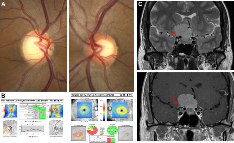
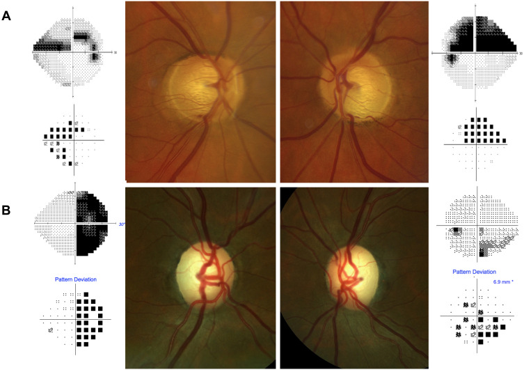
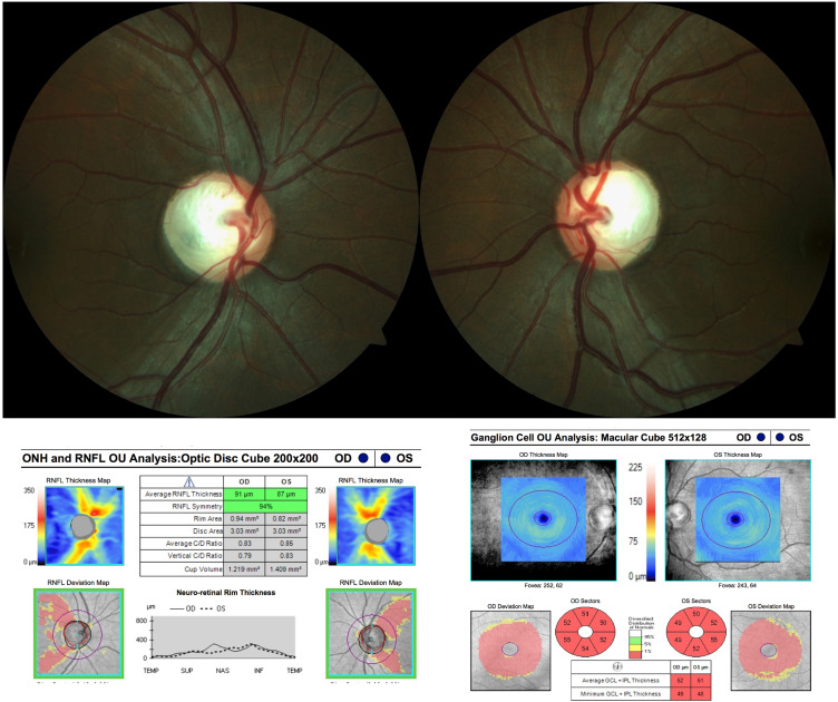
Optic nerve cupping or enlargement of the cup-to-disc ratio is widely recognized as a feature of glaucoma, however it may also occur in non-glaucomatous optic neuropathies. The most well-recognized non-glaucomatous optic neuropathies that cause cupping include compressive optic neuropathies, arteritic anterior ischemic optic neuropathies, hereditary optic neuropathies, and optic neuritis. Cupping is thought to consist of two main components: prelaminar and laminar thinning. The former is a shallow form of cupping and related to loss of retinal ganglion cells, whereas the latter involves damage to the lamina cribrosa and peripapillary scleral connective tissue. Differentiating glaucomatous and non-glaucomatous optic nerve cupping remains challenging even for experienced observers. Classically, the optic nerve in non-glaucomatous causes has pallor of the neuroretinal rim, but the optic nerve should not be examined in isolation. The patient's medical history, history of presenting illness, visual function (visual acuity, color vision and visual field testing) and ocular examination also need to be considered. Ancillary testing such as optical coherence tomography of the retinal nerve fiber layer and ganglion cell layer-inner plexiform layer may also be helpful in localizing the disease. In this review, we review the non-glaucomatous causes of cupping and provide an approach to evaluating a patient that presents with an enlarged cup-to-disc ratio.
期刊介绍:
Eye and Brain is an international, peer-reviewed, open access journal focusing on basic research, clinical findings, and expert reviews in the field of visual science and neuro-ophthalmology. The journal’s unique focus is the link between two well-known visual centres, the eye and the brain, with an emphasis on the importance of such connections. All aspects of clinical and especially basic research on the visual system are addressed within the journal as well as significant future directions in vision research and therapeutic measures. This unique journal focuses on neurological aspects of vision – both physiological and pathological. The scope of the journal spans from the cornea to the associational visual cortex and all the visual centers in between. Topics range from basic biological mechanisms to therapeutic treatment, from simple organisms to humans, and utilizing techniques from molecular biology to behavior. The journal especially welcomes primary research articles or review papers that make the connection between the eye and the brain. Specific areas covered in the journal include: Physiology and pathophysiology of visual centers, Eye movement disorders and strabismus, Cellular, biochemical, and molecular features of the visual system, Structural and functional organization of the eye and of the visual cortex, Metabolic demands of the visual system, Diseases and disorders with neuro-ophthalmic manifestations, Clinical and experimental neuro-ophthalmology and visual system pathologies, Epidemiological studies.


 分享
分享
 求助内容:
求助内容: 应助结果提醒方式:
 扫码关注我们
