Julia N Cheng, Jennifer B Frye, Susan A Whitman, Andrew G Kunihiro, Julia A Brickey, Janet L Funk
{"title":"Osteolytic effects of tumoral estrogen signaling in an estrogen receptor-positive breast cancer bone metastasis model.","authors":"Julia N Cheng, Jennifer B Frye, Susan A Whitman, Andrew G Kunihiro, Julia A Brickey, Janet L Funk","doi":"10.20517/2394-4722.2021.27","DOIUrl":null,"url":null,"abstract":"<p><strong>Aim: </strong>Estrogen receptor α-positive (ER+) subtypes of breast cancer have the greatest predilection for forming osteolytic bone metastases (BMETs). Because tumor-derived factors mediate osteolysis, a possible role for tumoral ERα signaling in driving ER+ BMET osteolysis was queried using an estrogen (E<sub>2</sub>)-dependent ER+ breast cancer BMET model.</p><p><strong>Methods: </strong>Female athymic Foxn1<sup>nu</sup> mice were inoculated with human ER+ MCF-7 breast cancer cells via the left cardiac ventricle post-E<sub>2</sub> pellet placement, and age- and dose-dependent E<sub>2</sub> effects on osteolytic ER+ BMET progression, as well as direct bone effects of E<sub>2</sub>, were determined.</p><p><strong>Results: </strong>Osteolytic BMETs, which did not form in the absence of E<sub>2</sub> supplementation, occurred with the same frequency in young (5-week-old) <i>vs.</i> skeletally mature (16-week-old) E<sub>2</sub> (0.72 mg)-treated mice, but were larger in young mice where anabolic bone effects of E<sub>2</sub> were greater. However, in mice of a single age and across a range of E<sub>2</sub> doses, anabolic E<sub>2</sub> bone effects were constant, while osteolytic ER+ BMET lesion incidence and size increased in an E<sub>2</sub>-dose-dependent fashion. Osteoclasts in ER+ tumor-bearing (but not tumor-naive) mice increased in an E<sub>2</sub>-dose dependent fashion at the bone-tumor interface, while histologic tumor size and proliferation did not vary with E<sub>2</sub> dose. E<sub>2</sub>-inducible tumoral secretion of the osteolytic factor parathyroid hormone-related protein (PTHrP) was dose-dependent and mediated by ERα, with significantly greater levels of secretion from ER+ BMET-derived tumor cells.</p><p><strong>Conclusion: </strong>These results suggest that tumoral ERα signaling may contribute to ER+ BMET-associated osteolysis, potentially explaining the greater predilection for ER+ tumors to form clinically-evident osteolytic BMETs.</p>","PeriodicalId":15167,"journal":{"name":"Journal of Cancer Metastasis and Treatment","volume":"7 ","pages":""},"PeriodicalIF":1.0000,"publicationDate":"2021-01-01","publicationTypes":"Journal Article","fieldsOfStudy":null,"isOpenAccess":false,"openAccessPdf":"https://www.ncbi.nlm.nih.gov/pmc/articles/PMC8594878/pdf/","citationCount":"0","resultStr":null,"platform":"Semanticscholar","paperid":null,"PeriodicalName":"Journal of Cancer Metastasis and Treatment","FirstCategoryId":"3","ListUrlMain":"https://doi.org/10.20517/2394-4722.2021.27","RegionNum":0,"RegionCategory":null,"ArticlePicture":[],"TitleCN":null,"AbstractTextCN":null,"PMCID":null,"EPubDate":"2021/4/8 0:00:00","PubModel":"Epub","JCR":"Q4","JCRName":"ONCOLOGY","Score":null,"Total":0}
引用次数: 0
Abstract
Aim: Estrogen receptor α-positive (ER+) subtypes of breast cancer have the greatest predilection for forming osteolytic bone metastases (BMETs). Because tumor-derived factors mediate osteolysis, a possible role for tumoral ERα signaling in driving ER+ BMET osteolysis was queried using an estrogen (E2)-dependent ER+ breast cancer BMET model.
Methods: Female athymic Foxn1nu mice were inoculated with human ER+ MCF-7 breast cancer cells via the left cardiac ventricle post-E2 pellet placement, and age- and dose-dependent E2 effects on osteolytic ER+ BMET progression, as well as direct bone effects of E2, were determined.
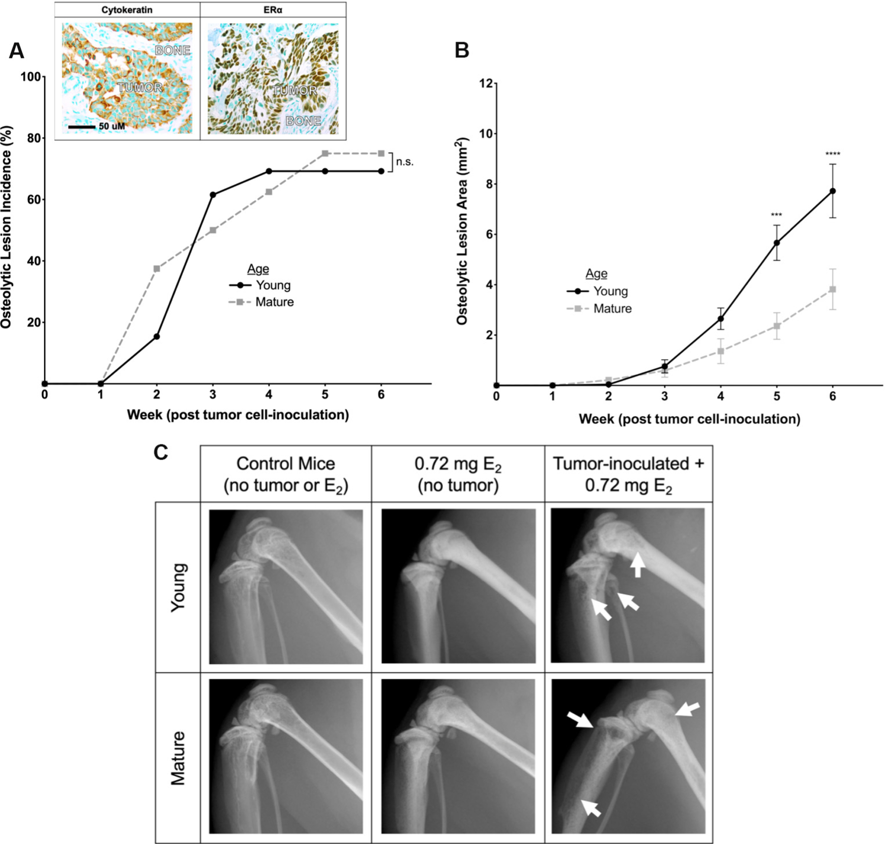
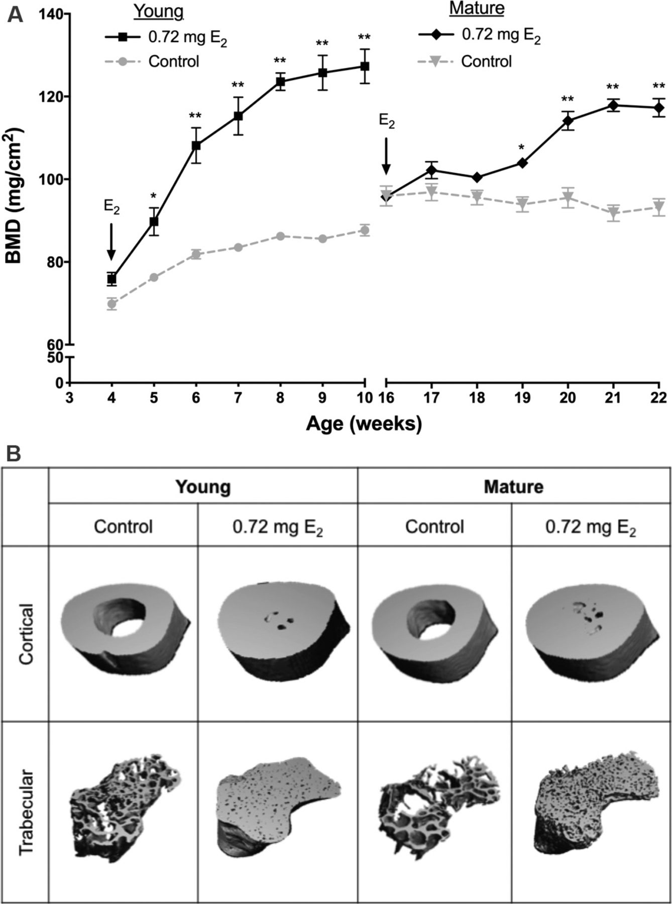
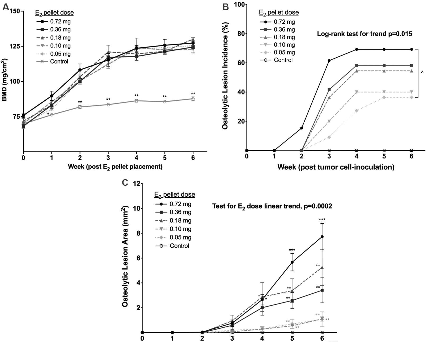
Results: Osteolytic BMETs, which did not form in the absence of E2 supplementation, occurred with the same frequency in young (5-week-old) vs. skeletally mature (16-week-old) E2 (0.72 mg)-treated mice, but were larger in young mice where anabolic bone effects of E2 were greater. However, in mice of a single age and across a range of E2 doses, anabolic E2 bone effects were constant, while osteolytic ER+ BMET lesion incidence and size increased in an E2-dose-dependent fashion. Osteoclasts in ER+ tumor-bearing (but not tumor-naive) mice increased in an E2-dose dependent fashion at the bone-tumor interface, while histologic tumor size and proliferation did not vary with E2 dose. E2-inducible tumoral secretion of the osteolytic factor parathyroid hormone-related protein (PTHrP) was dose-dependent and mediated by ERα, with significantly greater levels of secretion from ER+ BMET-derived tumor cells.
Conclusion: These results suggest that tumoral ERα signaling may contribute to ER+ BMET-associated osteolysis, potentially explaining the greater predilection for ER+ tumors to form clinically-evident osteolytic BMETs.


 分享
分享

 求助内容:
求助内容: 应助结果提醒方式:
 扫码关注我们
