{"title":"Super-resolution imaging to reveal the nanostructure of tripartite synapses.","authors":"Natalija Aleksejenko, Janosch P Heller","doi":"10.1042/NS20210003","DOIUrl":null,"url":null,"abstract":"<p><p>Even though neurons are the main drivers of information processing in the brain and spinal cord, other cell types are important to mediate adequate flow of information. These include electrically passive glial cells such as microglia and astrocytes, which recently emerged as active partners facilitating proper signal transduction. In disease, these cells undergo pathophysiological changes that propel disease progression and change synaptic connections and signal transmission. In the healthy brain, astrocytic processes contact pre- and postsynaptic structures. These processes can be nanoscopic, and therefore only electron microscopy has been able to reveal their structure and morphology. However, electron microscopy is not suitable in revealing dynamic changes, and it is labour- and time-intensive. The dawn of super-resolution microscopy, techniques that 'break' the diffraction limit of conventional light microscopy, over the last decades has enabled researchers to reveal the nanoscopic synaptic environment. In this review, we highlight and discuss recent advances in our understanding of the nano-world of the so-called tripartite synapses, the relationship between pre- and postsynapse as well as astrocytic processes. Overall, novel super-resolution microscopy methods are needed to fully illuminate the intimate relationship between glia and neuronal cells that underlies signal transduction in the brain and that might be affected in diseases such as Alzheimer's disease and epilepsy.</p>","PeriodicalId":74287,"journal":{"name":"Neuronal signaling","volume":"5 4","pages":"NS20210003"},"PeriodicalIF":0.0000,"publicationDate":"2021-10-14","publicationTypes":"Journal Article","fieldsOfStudy":null,"isOpenAccess":false,"openAccessPdf":"https://www.ncbi.nlm.nih.gov/pmc/articles/PMC8536832/pdf/","citationCount":"0","resultStr":null,"platform":"Semanticscholar","paperid":null,"PeriodicalName":"Neuronal signaling","FirstCategoryId":"1085","ListUrlMain":"https://doi.org/10.1042/NS20210003","RegionNum":0,"RegionCategory":null,"ArticlePicture":[],"TitleCN":null,"AbstractTextCN":null,"PMCID":null,"EPubDate":"2021/12/1 0:00:00","PubModel":"eCollection","JCR":"Q4","JCRName":"Neuroscience","Score":null,"Total":0}
引用次数: 0
Abstract
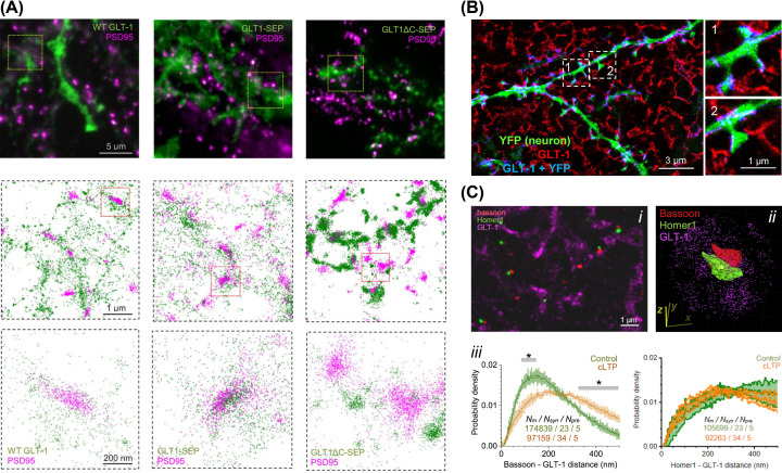
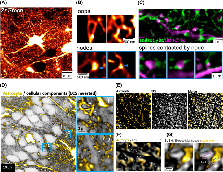
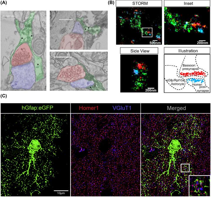
Even though neurons are the main drivers of information processing in the brain and spinal cord, other cell types are important to mediate adequate flow of information. These include electrically passive glial cells such as microglia and astrocytes, which recently emerged as active partners facilitating proper signal transduction. In disease, these cells undergo pathophysiological changes that propel disease progression and change synaptic connections and signal transmission. In the healthy brain, astrocytic processes contact pre- and postsynaptic structures. These processes can be nanoscopic, and therefore only electron microscopy has been able to reveal their structure and morphology. However, electron microscopy is not suitable in revealing dynamic changes, and it is labour- and time-intensive. The dawn of super-resolution microscopy, techniques that 'break' the diffraction limit of conventional light microscopy, over the last decades has enabled researchers to reveal the nanoscopic synaptic environment. In this review, we highlight and discuss recent advances in our understanding of the nano-world of the so-called tripartite synapses, the relationship between pre- and postsynapse as well as astrocytic processes. Overall, novel super-resolution microscopy methods are needed to fully illuminate the intimate relationship between glia and neuronal cells that underlies signal transduction in the brain and that might be affected in diseases such as Alzheimer's disease and epilepsy.


 分享
分享

 求助内容:
求助内容: 应助结果提醒方式:
 扫码关注我们
