Carol Vitellas, Ivo Besong Mangeb, Luis Regalado, Chiemezie Chianotu Amadi
{"title":"Mediastinal Extension of a Pancreatic Pseudocyst: A Rare Intrathoracic Complication of Pancreatitis.","authors":"Carol Vitellas, Ivo Besong Mangeb, Luis Regalado, Chiemezie Chianotu Amadi","doi":"10.1155/2021/1919550","DOIUrl":null,"url":null,"abstract":"<p><p>Pancreatic pseudocysts are a common complication of pancreatitis. However, mediastinal extension of a pseudocyst is rare and often presents with atypical symptoms. We present a case of mediastinal extension of a pancreatic pseudocyst in a 56-year-old woman with a history of alcohol-related chronic pancreatitis, who presented with acute on chronic epigastric abdominal pain and atypical chest pain. Serum lipase was elevated, and imaging by contrast-enhanced computed tomography (CT) demonstrated a paraesophageal fluid collection. This collection was continuous with a peripancreatic pseudocyst and extended into the posterior mediastinum via the esophageal hiatus. Mediastinal extension of a pancreatic pseudocyst was confirmed by magnetic resonance imaging (MRI). The patient was managed conservatively in the hospital with parenteral nutrition therapy, pain control, and close imaging observation. The patient was discharged home to continue conservative management and close imaging follow-up. An initial follow-up CT examination 8 weeks after discharge revealed interval decrease in the posterior mediastinal collection but also interval development of loculated left pleural and pericardial effusions.</p>","PeriodicalId":30326,"journal":{"name":"Case Reports in Radiology","volume":" ","pages":"1919550"},"PeriodicalIF":0.0000,"publicationDate":"2021-11-30","publicationTypes":"Journal Article","fieldsOfStudy":null,"isOpenAccess":false,"openAccessPdf":"https://www.ncbi.nlm.nih.gov/pmc/articles/PMC8651394/pdf/","citationCount":"2","resultStr":null,"platform":"Semanticscholar","paperid":null,"PeriodicalName":"Case Reports in Radiology","FirstCategoryId":"1085","ListUrlMain":"https://doi.org/10.1155/2021/1919550","RegionNum":0,"RegionCategory":null,"ArticlePicture":[],"TitleCN":null,"AbstractTextCN":null,"PMCID":null,"EPubDate":"2021/1/1 0:00:00","PubModel":"eCollection","JCR":"","JCRName":"","Score":null,"Total":0}
引用次数: 2
Abstract
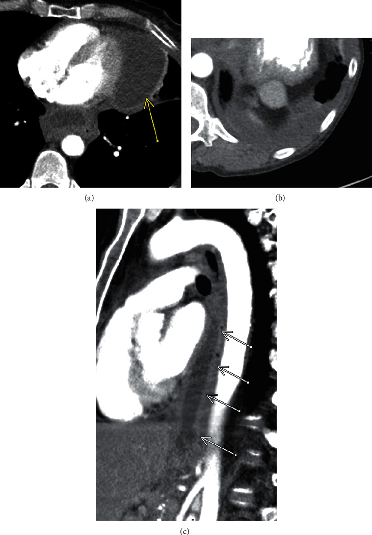
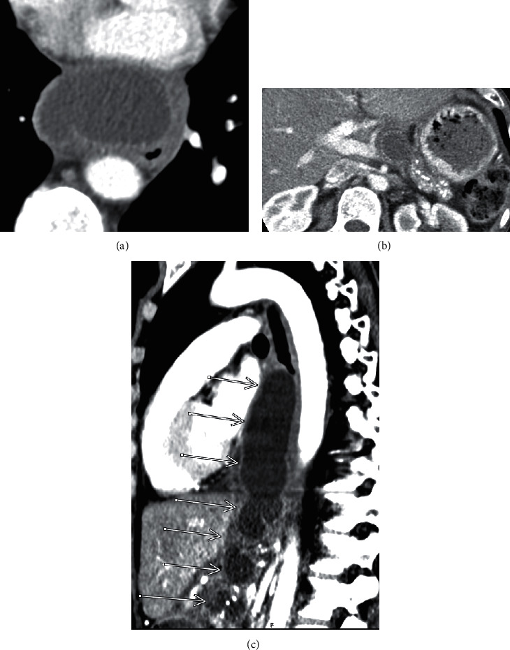
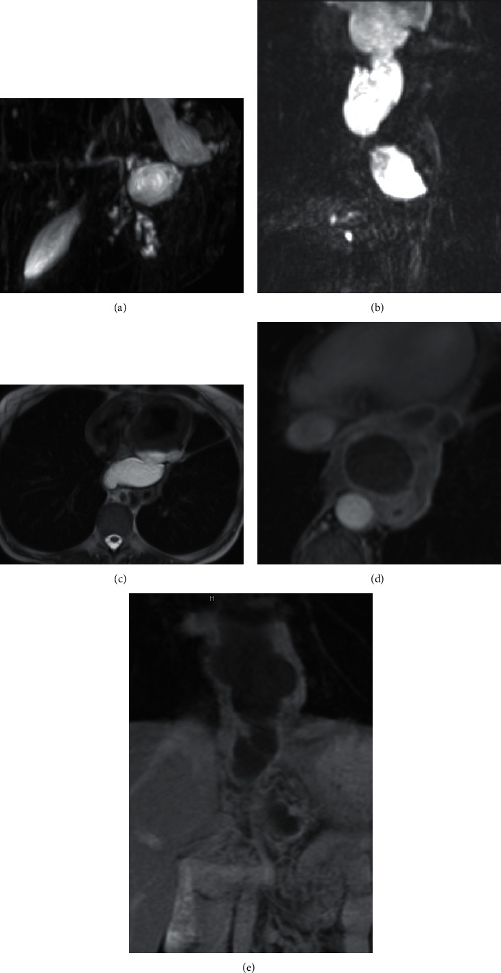
Pancreatic pseudocysts are a common complication of pancreatitis. However, mediastinal extension of a pseudocyst is rare and often presents with atypical symptoms. We present a case of mediastinal extension of a pancreatic pseudocyst in a 56-year-old woman with a history of alcohol-related chronic pancreatitis, who presented with acute on chronic epigastric abdominal pain and atypical chest pain. Serum lipase was elevated, and imaging by contrast-enhanced computed tomography (CT) demonstrated a paraesophageal fluid collection. This collection was continuous with a peripancreatic pseudocyst and extended into the posterior mediastinum via the esophageal hiatus. Mediastinal extension of a pancreatic pseudocyst was confirmed by magnetic resonance imaging (MRI). The patient was managed conservatively in the hospital with parenteral nutrition therapy, pain control, and close imaging observation. The patient was discharged home to continue conservative management and close imaging follow-up. An initial follow-up CT examination 8 weeks after discharge revealed interval decrease in the posterior mediastinal collection but also interval development of loculated left pleural and pericardial effusions.


 分享
分享

 求助内容:
求助内容: 应助结果提醒方式:
 扫码关注我们
