{"title":"Experimental Imaging Study of Encephalomalacia Fluid-Attenuated Inversion Recovery (FLAIR) Hyperintense Lesions in Posttraumatic Epilepsy.","authors":"Dan Wang, Kai Shang, Zheng Sun, Yue-Hua Li","doi":"10.1155/2021/2678379","DOIUrl":null,"url":null,"abstract":"<p><p>This study introduced new MRI techniques such as neurite orientation dispersion and density imaging (NODDI); NODDI applies a three-compartment tissue model to multishell DWI data that allows the examination of both the intra- and extracellular properties of white matter tissue. This, in turn, enables us to distinguish the two key aspects of axonal pathology-the packing density of axons in the white matter and the spatial organization of axons (orientation dispersion (OD)). NODDI is used to detect possible abnormalities of posttraumatic encephalomalacia fluid-attenuated inversion recovery (FLAIR) hyperintense lesions in neurite density and dispersion. <i>Methods</i>. 26 epilepsy patients associated with FLAIR hyperintensity around the trauma encephalomalacia region were in the epilepsy group. 18 posttraumatic patients with a FLAIR hyperintense encephalomalacia region were in the nonepilepsy group. Neurite density and dispersion affection in FLAIR hyperintense lesions around encephalomalacia were measured by NODDI using intracellular volume fraction (ICVF), and we compare these findings with conventional diffusion MRI parameters, namely, fractional anisotropy (FA) and apparent diffusion coefficient (ADC). Differences were compared between the epilepsy and nonepilepsy groups, as well as in the FLAIR hyperintense part and in the FLAIR hypointense part to try to find neurite density and dispersion differences in these parts. <i>Results</i>. ICVF of FLAIR hyperintense lesions in the epilepsy group was significantly higher than that in the nonepilepsy group (<i>P</i> < 0.001). ICVF reveals more information of FLAIR(+) and FLAIR(-) parts of encephalomalacia than OD and FA and ADC. <i>Conclusion</i>. The FLAIR hyperintense part around encephalomalacia in the epilepsy group showed higher ICVF, indicating that this part may have more neurite density and dispersion and may be contributing to epilepsy. NODDI indicated high neurite density with the intensity of myelin in the FLAIR hyperintense lesion. Therefore, NODDI likely shows that neurite density may be a more sensitive marker of pathology than FA.</p>","PeriodicalId":51299,"journal":{"name":"Neural Plasticity","volume":" ","pages":"2678379"},"PeriodicalIF":3.7000,"publicationDate":"2021-10-31","publicationTypes":"Journal Article","fieldsOfStudy":null,"isOpenAccess":false,"openAccessPdf":"https://www.ncbi.nlm.nih.gov/pmc/articles/PMC8572636/pdf/","citationCount":"2","resultStr":null,"platform":"Semanticscholar","paperid":null,"PeriodicalName":"Neural Plasticity","FirstCategoryId":"3","ListUrlMain":"https://doi.org/10.1155/2021/2678379","RegionNum":4,"RegionCategory":"医学","ArticlePicture":[],"TitleCN":null,"AbstractTextCN":null,"PMCID":null,"EPubDate":"2021/1/1 0:00:00","PubModel":"eCollection","JCR":"Q2","JCRName":"NEUROSCIENCES","Score":null,"Total":0}
引用次数: 2
Abstract
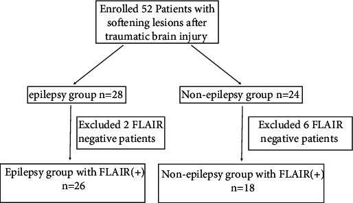
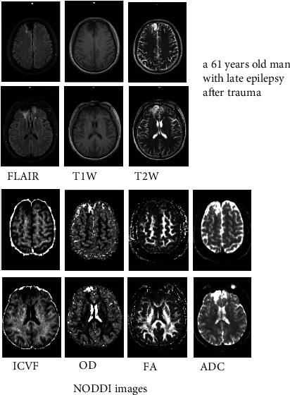
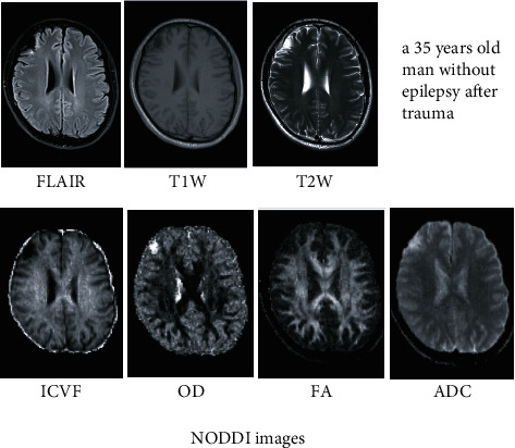
This study introduced new MRI techniques such as neurite orientation dispersion and density imaging (NODDI); NODDI applies a three-compartment tissue model to multishell DWI data that allows the examination of both the intra- and extracellular properties of white matter tissue. This, in turn, enables us to distinguish the two key aspects of axonal pathology-the packing density of axons in the white matter and the spatial organization of axons (orientation dispersion (OD)). NODDI is used to detect possible abnormalities of posttraumatic encephalomalacia fluid-attenuated inversion recovery (FLAIR) hyperintense lesions in neurite density and dispersion. Methods. 26 epilepsy patients associated with FLAIR hyperintensity around the trauma encephalomalacia region were in the epilepsy group. 18 posttraumatic patients with a FLAIR hyperintense encephalomalacia region were in the nonepilepsy group. Neurite density and dispersion affection in FLAIR hyperintense lesions around encephalomalacia were measured by NODDI using intracellular volume fraction (ICVF), and we compare these findings with conventional diffusion MRI parameters, namely, fractional anisotropy (FA) and apparent diffusion coefficient (ADC). Differences were compared between the epilepsy and nonepilepsy groups, as well as in the FLAIR hyperintense part and in the FLAIR hypointense part to try to find neurite density and dispersion differences in these parts. Results. ICVF of FLAIR hyperintense lesions in the epilepsy group was significantly higher than that in the nonepilepsy group (P < 0.001). ICVF reveals more information of FLAIR(+) and FLAIR(-) parts of encephalomalacia than OD and FA and ADC. Conclusion. The FLAIR hyperintense part around encephalomalacia in the epilepsy group showed higher ICVF, indicating that this part may have more neurite density and dispersion and may be contributing to epilepsy. NODDI indicated high neurite density with the intensity of myelin in the FLAIR hyperintense lesion. Therefore, NODDI likely shows that neurite density may be a more sensitive marker of pathology than FA.
期刊介绍:
Neural Plasticity is an international, interdisciplinary journal dedicated to the publication of articles related to all aspects of neural plasticity, with special emphasis on its functional significance as reflected in behavior and in psychopathology. Neural Plasticity publishes research and review articles from the entire range of relevant disciplines, including basic neuroscience, behavioral neuroscience, cognitive neuroscience, biological psychology, and biological psychiatry.


 分享
分享
 求助内容:
求助内容: 应助结果提醒方式:
 扫码关注我们
