Mohammed H. AL Mughram , Noah B. Herrington , Claudio Catalano , Glen E. Kellogg
{"title":"Systematized analysis of secondary structure dependence of key structural features of residues in soluble and membrane-bound proteins","authors":"Mohammed H. AL Mughram , Noah B. Herrington , Claudio Catalano , Glen E. Kellogg","doi":"10.1016/j.yjsbx.2021.100055","DOIUrl":null,"url":null,"abstract":"<div><p>Knowledge of three-dimensional protein structure is integral to most modern drug discovery efforts. Recent advancements have highlighted new techniques for 3D protein structure determination and, where structural data cannot be collected experimentally, prediction of protein structure. We have undertaken a major effort to use existing protein structures to collect, characterize, and catalogue the inter-atomic interactions that define and compose 3D structure by mapping hydropathic interaction environments as maps in 3D space. This work has been performed on a residue-by-residue basis, where we have seen evidence for relationships between environment character, residue solvent-accessible surface areas and their secondary structures. In this graphical review, we apply principles from our earlier studies and expand the scope to all common amino acid residue types in both soluble and membrane proteins. Key to this analysis is parsing the Ramachandran plot to an 8-by-8 chessboard to define secondary structure bins. Our analysis yielded a number of quantitative discoveries: 1) increased fraction of hydrophobic residues (alanine, isoleucine, leucine, phenylalanine and valine) in membrane proteins compared to their fractions in soluble proteins; 2) less burial coupled with significant increases in favorable hydrophobic interactions for hydrophobic residues in membrane proteins compared to soluble proteins; and 3) higher burial and more favorable polar interactions for polar residues now preferring the interior of membrane proteins. These observations and the supporting data should provide benchmarks for current studies of protein residues in different environments and may be able to guide future protein structure prediction efforts.</p></div>","PeriodicalId":17238,"journal":{"name":"Journal of Structural Biology: X","volume":"5 ","pages":"Article 100055"},"PeriodicalIF":5.1000,"publicationDate":"2021-01-01","publicationTypes":"Journal Article","fieldsOfStudy":null,"isOpenAccess":false,"openAccessPdf":"https://ftp.ncbi.nlm.nih.gov/pub/pmc/oa_pdf/33/90/main.PMC8654985.pdf","citationCount":"2","resultStr":null,"platform":"Semanticscholar","paperid":null,"PeriodicalName":"Journal of Structural Biology: X","FirstCategoryId":"1085","ListUrlMain":"https://www.sciencedirect.com/science/article/pii/S259015242100012X","RegionNum":0,"RegionCategory":null,"ArticlePicture":[],"TitleCN":null,"AbstractTextCN":null,"PMCID":null,"EPubDate":"","PubModel":"","JCR":"Q2","JCRName":"BIOCHEMISTRY & MOLECULAR BIOLOGY","Score":null,"Total":0}
引用次数: 2
Abstract
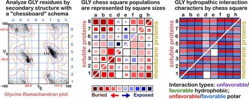
Knowledge of three-dimensional protein structure is integral to most modern drug discovery efforts. Recent advancements have highlighted new techniques for 3D protein structure determination and, where structural data cannot be collected experimentally, prediction of protein structure. We have undertaken a major effort to use existing protein structures to collect, characterize, and catalogue the inter-atomic interactions that define and compose 3D structure by mapping hydropathic interaction environments as maps in 3D space. This work has been performed on a residue-by-residue basis, where we have seen evidence for relationships between environment character, residue solvent-accessible surface areas and their secondary structures. In this graphical review, we apply principles from our earlier studies and expand the scope to all common amino acid residue types in both soluble and membrane proteins. Key to this analysis is parsing the Ramachandran plot to an 8-by-8 chessboard to define secondary structure bins. Our analysis yielded a number of quantitative discoveries: 1) increased fraction of hydrophobic residues (alanine, isoleucine, leucine, phenylalanine and valine) in membrane proteins compared to their fractions in soluble proteins; 2) less burial coupled with significant increases in favorable hydrophobic interactions for hydrophobic residues in membrane proteins compared to soluble proteins; and 3) higher burial and more favorable polar interactions for polar residues now preferring the interior of membrane proteins. These observations and the supporting data should provide benchmarks for current studies of protein residues in different environments and may be able to guide future protein structure prediction efforts.


 分享
分享
 求助内容:
求助内容: 应助结果提醒方式:
 扫码关注我们
