Assessment of the Bone Mineral Density and Microstructure of the Human Femoral Head according to Different Tip-apex Distances Can Guide the Treatment of Intertrochanteric Hip Fractures.
{"title":"Assessment of the Bone Mineral Density and Microstructure of the Human Femoral Head according to Different Tip-apex Distances Can Guide the Treatment of Intertrochanteric Hip Fractures.","authors":"Quan-Hu Shen, JiWoong Baik, YeYeon Won","doi":"10.5371/hp.2021.33.4.190","DOIUrl":null,"url":null,"abstract":"<p><strong>Purpose: </strong>We analyzed the microstructure and bone mineral density (BMD) of the trabecular bone in the femoral head of patients with osteoporosis.</p><p><strong>Materials and methods: </strong>Sixteen femoral heads with osteoporotic femoral neck fractures underwent micro-computed tomography scanning. In each tip-apex distance (TAD) of 15, 20, and 25 mm, five regions of interest (ROIs) were extracted from the central, anterior, posterior, superior, and inferior sections. A total of 15 ROIs were extracted from TADs of 15, 20, and 25 mm. The measurement parameters included BMD, percent bone volume: bone volume/total volume (BV/TV), trabecular thickness (Tb.Th), trabecular number (Tb.N), structural model index (SMI), and degree of anisotropy (DOA).</p><p><strong>Results: </strong>The lowest BMD and BV/TV values were observed in the inferior region and differed significantly from those in other regions (<i>P</i><0.05). Lower Tb.Th and Tb.N values were observed in the inferior region compared with those in the central region (<i>P</i><0.05). The highest SMI value was observed in the inferior region (<i>P</i><0.05). With TAD of 15 and 20 mm, the DOA values in the inferior region were lower than those in the anterior region (<i>P</i><0.05). Lower BMD and BV/TV values were observed in the anterior, central, and inferior regions of TAD of 15 mm compared with those in the corresponding regions of TAD of 25 mm (<i>P</i><0.05).</p><p><strong>Conclusion: </strong>Positioning the lag screw between TAD of 20 to 25 mm and in the inferior region is recommended, and TAD of less than 15 mm is not recommended.</p>","PeriodicalId":73239,"journal":{"name":"Hip & pelvis","volume":"33 4","pages":"190-199"},"PeriodicalIF":0.0000,"publicationDate":"2021-12-01","publicationTypes":"Journal Article","fieldsOfStudy":null,"isOpenAccess":false,"openAccessPdf":"https://ftp.ncbi.nlm.nih.gov/pub/pmc/oa_pdf/f6/f5/hp-33-190.PMC8654587.pdf","citationCount":"2","resultStr":null,"platform":"Semanticscholar","paperid":null,"PeriodicalName":"Hip & pelvis","FirstCategoryId":"1085","ListUrlMain":"https://doi.org/10.5371/hp.2021.33.4.190","RegionNum":0,"RegionCategory":null,"ArticlePicture":[],"TitleCN":null,"AbstractTextCN":null,"PMCID":null,"EPubDate":"","PubModel":"","JCR":"","JCRName":"","Score":null,"Total":0}
引用次数: 2
Abstract
Purpose: We analyzed the microstructure and bone mineral density (BMD) of the trabecular bone in the femoral head of patients with osteoporosis.
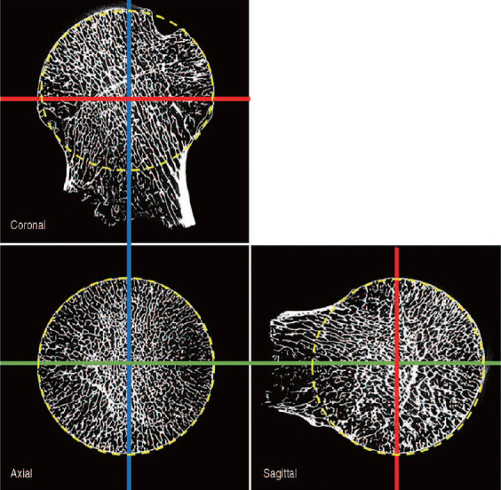
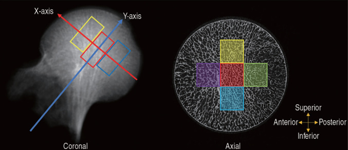
Materials and methods: Sixteen femoral heads with osteoporotic femoral neck fractures underwent micro-computed tomography scanning. In each tip-apex distance (TAD) of 15, 20, and 25 mm, five regions of interest (ROIs) were extracted from the central, anterior, posterior, superior, and inferior sections. A total of 15 ROIs were extracted from TADs of 15, 20, and 25 mm. The measurement parameters included BMD, percent bone volume: bone volume/total volume (BV/TV), trabecular thickness (Tb.Th), trabecular number (Tb.N), structural model index (SMI), and degree of anisotropy (DOA).
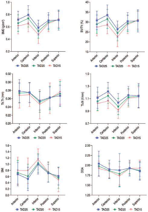
Results: The lowest BMD and BV/TV values were observed in the inferior region and differed significantly from those in other regions (P<0.05). Lower Tb.Th and Tb.N values were observed in the inferior region compared with those in the central region (P<0.05). The highest SMI value was observed in the inferior region (P<0.05). With TAD of 15 and 20 mm, the DOA values in the inferior region were lower than those in the anterior region (P<0.05). Lower BMD and BV/TV values were observed in the anterior, central, and inferior regions of TAD of 15 mm compared with those in the corresponding regions of TAD of 25 mm (P<0.05).
Conclusion: Positioning the lag screw between TAD of 20 to 25 mm and in the inferior region is recommended, and TAD of less than 15 mm is not recommended.


 分享
分享

 求助内容:
求助内容: 应助结果提醒方式:
 扫码关注我们
