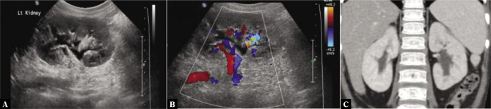
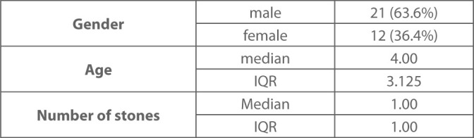
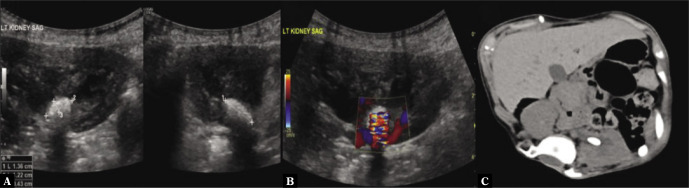
{"title":"Twinkle artifact in renal ultrasound, is it a solid point for the diagnosis of renal stone in children?","authors":"Moath AlSaiady, Ahmad Alqatie, Musab Almushayqih","doi":"10.15557/JoU.2021.0048","DOIUrl":null,"url":null,"abstract":"<p><p><b>Background:</b> Twinkle artifact, also known as color Doppler comet-tail artifact, occurs behind very strong, granular, and irregular reflecting interfaces such as crystals, stones, or calcification. This is visualized as a random mixture of red and blue pixels in the high-frequency shift spectrum located deep to the interface. Study results have suggested that the sonographic twinkling artifact may aid in the detection of renal stones with a variety of reference standard imaging modalities, including abdominal radiography, excretory urography, gray-scale sonography, and CT. <b>Material and methods:</b> Our retrospective observational study included children who had undergone abdomen/renal ultrasound for kidneys stones in our radiology department between 2013 and 2019. Presence of the twinkle artifact, and stone numbers and sizes were documented. CT examinations done <3 months prior to or after US were retrospectively assessed to confirm the presence of kidney stones as a reference standard. <b>Results:</b> Thirty-three abdominal renal US scans of 33 patients (21 males, 12 females) fulfilled the entry criteria. The interval between the US and CT was <3 months for all patients. The median overall age of the patients was 4 years (IQR: 3.125, range: 1- 165 months), The median number of days between the US and CT was 13 (IQR: 26, range: 0-81 days). US detected 33 hyperechoic foci suspected to be stones; 26 were confirmed as true positive (i.e. showed the twinkle artifact and were seen in CT), 4 were false positive (showed the twinkle artifact but were not seen in CT), and 3 were false negative (did not show the twinkle artifact but were seen in CT). The overall median stone size was 2 mm in the right kidney, and 5 mm in the left kidney (IQR: 6,11 mm), respectively. Twinkle artifact sensitivity was found to be 89.7% (95% CI 39.574%-90%). The twinkle artifact was assessed in all true-positive stones, determining a relatively high PPV of 26/29 (86.7%) for the twinkle artifact. The twinkle artifact was not dependent on stone size. Specificity for the twinkle artifact could not be calculated due to a lack of true negatives. <b>Conclusion:</b> The twinkle artifact is a sensitive US tool for detecting pediatric kidney and ureter stones, but with a small risk of false positive findings.</p>","PeriodicalId":45612,"journal":{"name":"Journal of Ultrasonography","volume":"21 87","pages":"e282-e285"},"PeriodicalIF":1.5000,"publicationDate":"2021-11-29","publicationTypes":"Journal Article","fieldsOfStudy":null,"isOpenAccess":false,"openAccessPdf":"https://ftp.ncbi.nlm.nih.gov/pub/pmc/oa_pdf/17/53/jou-21-87-e282.PMC8678643.pdf","citationCount":"2","resultStr":null,"platform":"Semanticscholar","paperid":null,"PeriodicalName":"Journal of Ultrasonography","FirstCategoryId":"1085","ListUrlMain":"https://doi.org/10.15557/JoU.2021.0048","RegionNum":0,"RegionCategory":null,"ArticlePicture":[],"TitleCN":null,"AbstractTextCN":null,"PMCID":null,"EPubDate":"2021/12/15 0:00:00","PubModel":"Epub","JCR":"Q3","JCRName":"RADIOLOGY, NUCLEAR MEDICINE & MEDICAL IMAGING","Score":null,"Total":0}
引用次数: 2
Abstract
Background: Twinkle artifact, also known as color Doppler comet-tail artifact, occurs behind very strong, granular, and irregular reflecting interfaces such as crystals, stones, or calcification. This is visualized as a random mixture of red and blue pixels in the high-frequency shift spectrum located deep to the interface. Study results have suggested that the sonographic twinkling artifact may aid in the detection of renal stones with a variety of reference standard imaging modalities, including abdominal radiography, excretory urography, gray-scale sonography, and CT. Material and methods: Our retrospective observational study included children who had undergone abdomen/renal ultrasound for kidneys stones in our radiology department between 2013 and 2019. Presence of the twinkle artifact, and stone numbers and sizes were documented. CT examinations done <3 months prior to or after US were retrospectively assessed to confirm the presence of kidney stones as a reference standard. Results: Thirty-three abdominal renal US scans of 33 patients (21 males, 12 females) fulfilled the entry criteria. The interval between the US and CT was <3 months for all patients. The median overall age of the patients was 4 years (IQR: 3.125, range: 1- 165 months), The median number of days between the US and CT was 13 (IQR: 26, range: 0-81 days). US detected 33 hyperechoic foci suspected to be stones; 26 were confirmed as true positive (i.e. showed the twinkle artifact and were seen in CT), 4 were false positive (showed the twinkle artifact but were not seen in CT), and 3 were false negative (did not show the twinkle artifact but were seen in CT). The overall median stone size was 2 mm in the right kidney, and 5 mm in the left kidney (IQR: 6,11 mm), respectively. Twinkle artifact sensitivity was found to be 89.7% (95% CI 39.574%-90%). The twinkle artifact was assessed in all true-positive stones, determining a relatively high PPV of 26/29 (86.7%) for the twinkle artifact. The twinkle artifact was not dependent on stone size. Specificity for the twinkle artifact could not be calculated due to a lack of true negatives. Conclusion: The twinkle artifact is a sensitive US tool for detecting pediatric kidney and ureter stones, but with a small risk of false positive findings.


 分享
分享
 求助内容:
求助内容: 应助结果提醒方式:
 扫码关注我们
