{"title":"Asymmetrical organization of oral structures in the primary and secondary somatosensory cortices in rats: An optical imaging study.","authors":"Yuki Kirihara, Manabu Zama, Satoshi Fujita, Shouhei Ogisawa, Shuichi Nishikubo, Morio Tonogi, Masayuki Kobayashi","doi":"10.1002/syn.22222","DOIUrl":null,"url":null,"abstract":"<p><p>In rodents, the representation of the body surface in the primary somatosensory cortex (S1) forms a mirror image along the ventral border of the S1 in the secondary somatosensory cortex (S2). Sensory information from the oral region is processed in the S1 and the border region between the S2 and insular oral region (IOR). We examined the relationship between somatosensory representations in the S1 and S2/IOR using optical imaging with a voltage-sensitive dye in urethane-anesthetized rats. In reference to the rhinal fissure and middle cerebral artery, we made a somatosensory map by applying electrical or air puff stimulation. The initial neural excitation in the S1 to facial structures, including the eyebrow, cornea, pinna, whisker pad, nasal tip, and nasal mucosa, spread toward the ventral area, putatively the S2. The initial cortical responses in the S1 to oral structures, including the lower lip, tongue, and teeth, were spatially separated from those in the S2/IOR. The representation of the tongue center, tongue tip, mandibular molar pulp, mandibular incisor pulp, and mandibular incisor periodontal ligament were almost linearly arranged from caudal to rostral in both S1 and S2/IOR. The lower lip was represented in the dorsal area from the representation of teeth and tongue in both S1 and S2/IOR. The representations of maxillary teeth were caudal and dorsal to the representations of mandibular teeth in the S1 and S2/IOR, respectively. These results suggest that the representation of oral structures in the S1 formed a non-mirror image, not a mirror image, in the S2/IOR.</p>","PeriodicalId":22131,"journal":{"name":"Synapse","volume":"76 1-2","pages":"e22222"},"PeriodicalIF":2.0000,"publicationDate":"2022-02-01","publicationTypes":"Journal Article","fieldsOfStudy":null,"isOpenAccess":false,"openAccessPdf":"","citationCount":"1","resultStr":null,"platform":"Semanticscholar","paperid":null,"PeriodicalName":"Synapse","FirstCategoryId":"3","ListUrlMain":"https://doi.org/10.1002/syn.22222","RegionNum":4,"RegionCategory":"医学","ArticlePicture":[],"TitleCN":null,"AbstractTextCN":null,"PMCID":null,"EPubDate":"2022/1/23 0:00:00","PubModel":"Epub","JCR":"Q4","JCRName":"NEUROSCIENCES","Score":null,"Total":0}
引用次数: 1
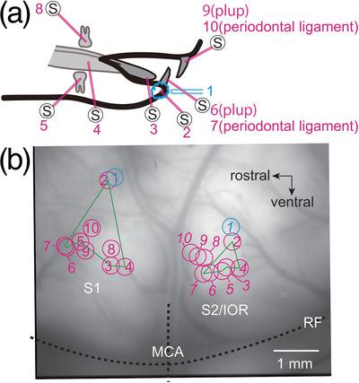
Abstract
In rodents, the representation of the body surface in the primary somatosensory cortex (S1) forms a mirror image along the ventral border of the S1 in the secondary somatosensory cortex (S2). Sensory information from the oral region is processed in the S1 and the border region between the S2 and insular oral region (IOR). We examined the relationship between somatosensory representations in the S1 and S2/IOR using optical imaging with a voltage-sensitive dye in urethane-anesthetized rats. In reference to the rhinal fissure and middle cerebral artery, we made a somatosensory map by applying electrical or air puff stimulation. The initial neural excitation in the S1 to facial structures, including the eyebrow, cornea, pinna, whisker pad, nasal tip, and nasal mucosa, spread toward the ventral area, putatively the S2. The initial cortical responses in the S1 to oral structures, including the lower lip, tongue, and teeth, were spatially separated from those in the S2/IOR. The representation of the tongue center, tongue tip, mandibular molar pulp, mandibular incisor pulp, and mandibular incisor periodontal ligament were almost linearly arranged from caudal to rostral in both S1 and S2/IOR. The lower lip was represented in the dorsal area from the representation of teeth and tongue in both S1 and S2/IOR. The representations of maxillary teeth were caudal and dorsal to the representations of mandibular teeth in the S1 and S2/IOR, respectively. These results suggest that the representation of oral structures in the S1 formed a non-mirror image, not a mirror image, in the S2/IOR.
期刊介绍:
SYNAPSE publishes articles concerned with all aspects of synaptic structure and function. This includes neurotransmitters, neuropeptides, neuromodulators, receptors, gap junctions, metabolism, plasticity, circuitry, mathematical modeling, ion channels, patch recording, single unit recording, development, behavior, pathology, toxicology, etc.


 分享
分享
 求助内容:
求助内容: 应助结果提醒方式:
 扫码关注我们
