Jing Han, Xi Yang, Wei Xu, Ronghua Jin, Weiyuan Liu, Lei Ding, Sha Meng, Yuan Zhang, Jin Li, Ying Zheng, Haowen Li, Fankun Meng
{"title":"Pilot study on the value of echocardiography combined with lung ultrasound to evaluate COVID-19 pneumonia.","authors":"Jing Han, Xi Yang, Wei Xu, Ronghua Jin, Weiyuan Liu, Lei Ding, Sha Meng, Yuan Zhang, Jin Li, Ying Zheng, Haowen Li, Fankun Meng","doi":"10.1186/s12947-021-00271-0","DOIUrl":null,"url":null,"abstract":"<p><strong>Background: </strong>This study aimed to investigate the relationship between echocardiography results and lung ultrasound score (LUS) in coronavirus disease 2019 (COVID-19) pneumonia patients and evaluate the impact of the combined application of these techniques in the evaluation of COVID-19 pneumonia.</p><p><strong>Methods: </strong>Hospitalized COVID-19 pneumonia patients who underwent daily lung ultrasound and echocardiography were included in this study. Patients with tricuspid regurgitation within three days of admission were enrolled. Moreover, the correlation and differences between their pulmonary artery pressure (PAP) and LUS on days 3, 8, and 13 were analyzed. The inner diameter of the pulmonary artery root as well as the size of the atria and ventricles were also considered.</p><p><strong>Results: </strong>The PAP on days 3, 8, and 13 of hospitalization was positively correlated with the LUS (r = 0.448, p = 0.003; r = 0.738, p < 0.001; r = 0.325, p = 0.036, respectively). On day 8, the values of both PAP and LUS were higher than on days 3 and 13 (p < 0.01). Similarly, PAP and LUS were significantly increased in 92.9% (39/42) and 90.5% (38/42) of patients, respectively, and at least one of these two values was positive in 97.6% (41/42) of cases. The inner diameters of the right atrium, right ventricle, and pulmonary artery also differed significantly from their corresponding values on days 3 and 13 (p < 0.05).</p><p><strong>Conclusions: </strong>PAP is positively correlated with LUS in COVID-19 pneumonia. The two values could be combined for a more precise assessment of disease progression and recovery status.</p>","PeriodicalId":9613,"journal":{"name":"Cardiovascular Ultrasound","volume":" ","pages":"2"},"PeriodicalIF":1.6000,"publicationDate":"2022-01-19","publicationTypes":"Journal Article","fieldsOfStudy":null,"isOpenAccess":false,"openAccessPdf":"https://www.ncbi.nlm.nih.gov/pmc/articles/PMC8767359/pdf/","citationCount":"0","resultStr":null,"platform":"Semanticscholar","paperid":null,"PeriodicalName":"Cardiovascular Ultrasound","FirstCategoryId":"3","ListUrlMain":"https://doi.org/10.1186/s12947-021-00271-0","RegionNum":3,"RegionCategory":"医学","ArticlePicture":[],"TitleCN":null,"AbstractTextCN":null,"PMCID":null,"EPubDate":"","PubModel":"","JCR":"Q3","JCRName":"CARDIAC & CARDIOVASCULAR SYSTEMS","Score":null,"Total":0}
引用次数: 0
Abstract
Background: This study aimed to investigate the relationship between echocardiography results and lung ultrasound score (LUS) in coronavirus disease 2019 (COVID-19) pneumonia patients and evaluate the impact of the combined application of these techniques in the evaluation of COVID-19 pneumonia.
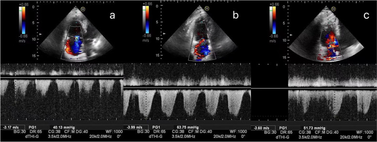
Methods: Hospitalized COVID-19 pneumonia patients who underwent daily lung ultrasound and echocardiography were included in this study. Patients with tricuspid regurgitation within three days of admission were enrolled. Moreover, the correlation and differences between their pulmonary artery pressure (PAP) and LUS on days 3, 8, and 13 were analyzed. The inner diameter of the pulmonary artery root as well as the size of the atria and ventricles were also considered.
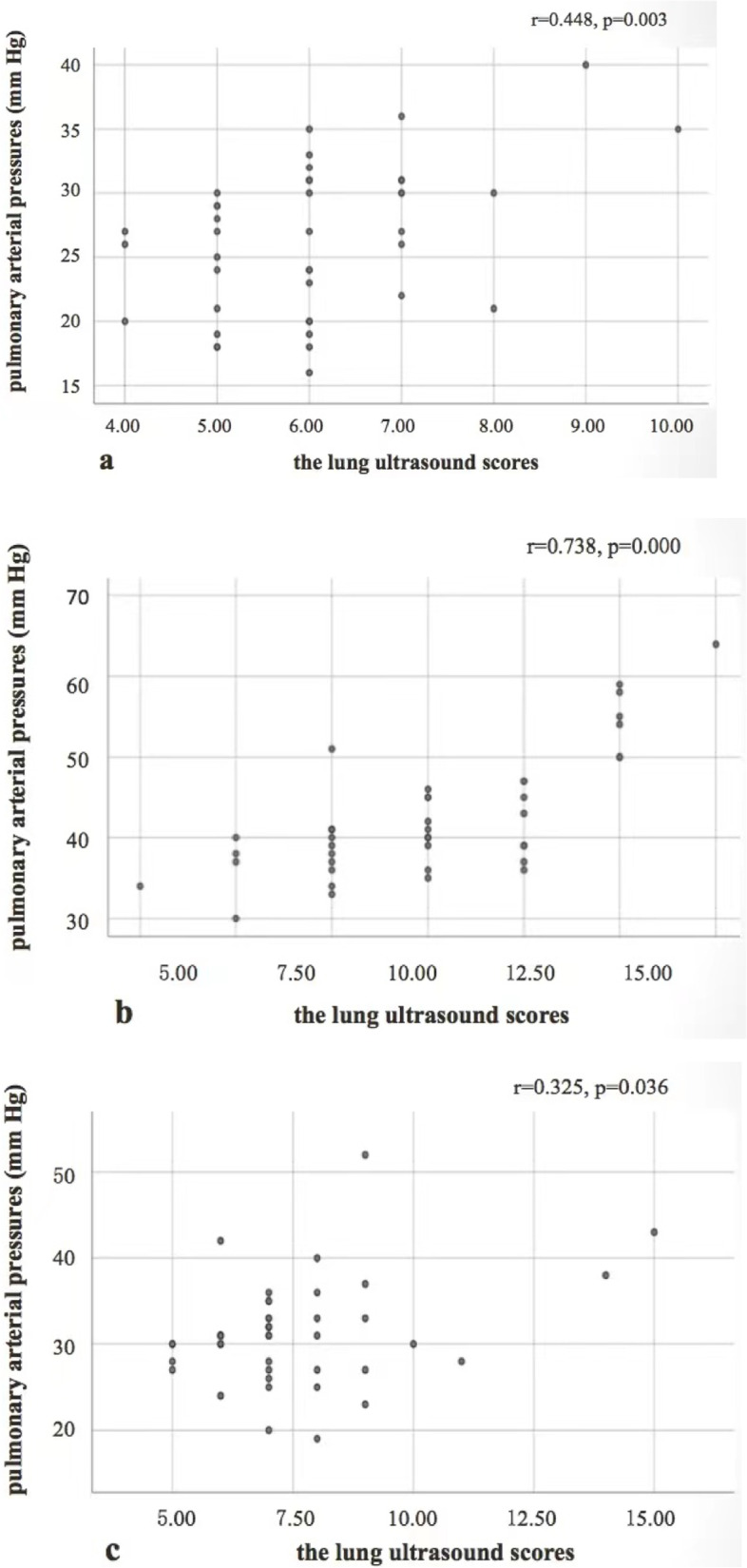
Results: The PAP on days 3, 8, and 13 of hospitalization was positively correlated with the LUS (r = 0.448, p = 0.003; r = 0.738, p < 0.001; r = 0.325, p = 0.036, respectively). On day 8, the values of both PAP and LUS were higher than on days 3 and 13 (p < 0.01). Similarly, PAP and LUS were significantly increased in 92.9% (39/42) and 90.5% (38/42) of patients, respectively, and at least one of these two values was positive in 97.6% (41/42) of cases. The inner diameters of the right atrium, right ventricle, and pulmonary artery also differed significantly from their corresponding values on days 3 and 13 (p < 0.05).
Conclusions: PAP is positively correlated with LUS in COVID-19 pneumonia. The two values could be combined for a more precise assessment of disease progression and recovery status.
期刊介绍:
Cardiovascular Ultrasound is an online journal, publishing peer-reviewed: original research; authoritative reviews; case reports on challenging and/or unusual diagnostic aspects; and expert opinions on new techniques and technologies. We are particularly interested in articles that include relevant images or video files, which provide an additional dimension to published articles and enhance understanding.
As an open access journal, Cardiovascular Ultrasound ensures high visibility for authors in addition to providing an up-to-date and freely available resource for the community. The journal welcomes discussion, and provides a forum for publishing opinion and debate ranging from biology to engineering to clinical echocardiography, with both speed and versatility.


 分享
分享
 求助内容:
求助内容: 应助结果提醒方式:
 扫码关注我们
