Zohaib Y Ahmad, Luis E Diaz, Frank W Roemer, Ajay Goud, Ali Guermazi
{"title":"Imaging Review of Subscapularis Tendon and Rotator Interval Pathology.","authors":"Zohaib Y Ahmad, Luis E Diaz, Frank W Roemer, Ajay Goud, Ali Guermazi","doi":"10.1155/2022/4009829","DOIUrl":null,"url":null,"abstract":"<p><p>As the largest rotator cuff muscle, the subscapularis plays a major role in stabilizing the glenohumeral joint, in conjunction with surrounding rotator cuff structures. Injury to the subscapularis tendon can be isolated, but more commonly is seen in conjunction with supraspinatus tendon pathology. Injury can be associated with biceps pulley instability, superior labral anterior-posterior (SLAP) tears, humeral head subluxation, and anterosuperior and coracoid impingements. The involvement of the rotator interval can lead to what is called \"the hidden lesion,\" due to its difficulty to diagnose during arthroscopy. Understanding the anatomical relations of the subscapularis tendon with the rest of the rotator cuff and rotator interval, as well as common patterns of injury that involve the subscapularis tendon, can aid in proper diagnosis of these injuries leading to prompt surgical repair. This review describes the anatomy of the subscapularis muscle and tendon, and the magnetic resonance imaging (MRI) patterns of subscapularis tendon injury.</p>","PeriodicalId":51864,"journal":{"name":"Radiology Research and Practice","volume":" ","pages":"4009829"},"PeriodicalIF":1.5000,"publicationDate":"2022-01-11","publicationTypes":"Journal Article","fieldsOfStudy":null,"isOpenAccess":false,"openAccessPdf":"https://www.ncbi.nlm.nih.gov/pmc/articles/PMC8767392/pdf/","citationCount":"4","resultStr":null,"platform":"Semanticscholar","paperid":null,"PeriodicalName":"Radiology Research and Practice","FirstCategoryId":"1085","ListUrlMain":"https://doi.org/10.1155/2022/4009829","RegionNum":0,"RegionCategory":null,"ArticlePicture":[],"TitleCN":null,"AbstractTextCN":null,"PMCID":null,"EPubDate":"2022/1/1 0:00:00","PubModel":"eCollection","JCR":"Q2","JCRName":"RADIOLOGY, NUCLEAR MEDICINE & MEDICAL IMAGING","Score":null,"Total":0}
引用次数: 4
Abstract
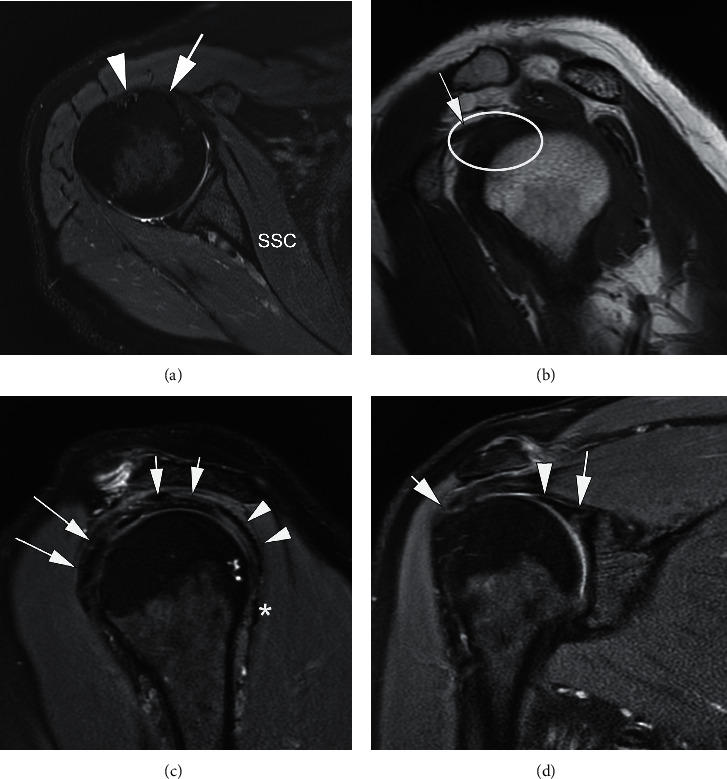
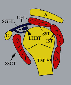
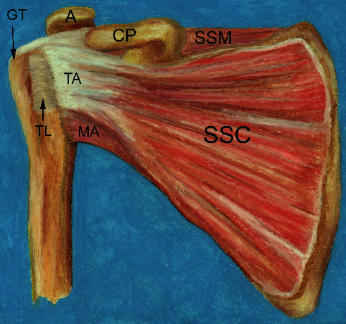
As the largest rotator cuff muscle, the subscapularis plays a major role in stabilizing the glenohumeral joint, in conjunction with surrounding rotator cuff structures. Injury to the subscapularis tendon can be isolated, but more commonly is seen in conjunction with supraspinatus tendon pathology. Injury can be associated with biceps pulley instability, superior labral anterior-posterior (SLAP) tears, humeral head subluxation, and anterosuperior and coracoid impingements. The involvement of the rotator interval can lead to what is called "the hidden lesion," due to its difficulty to diagnose during arthroscopy. Understanding the anatomical relations of the subscapularis tendon with the rest of the rotator cuff and rotator interval, as well as common patterns of injury that involve the subscapularis tendon, can aid in proper diagnosis of these injuries leading to prompt surgical repair. This review describes the anatomy of the subscapularis muscle and tendon, and the magnetic resonance imaging (MRI) patterns of subscapularis tendon injury.
期刊介绍:
Radiology Research and Practice is a peer-reviewed, Open Access journal that publishes articles on all areas of medical imaging. The journal promotes evidence-based radiology practice though the publication of original research, reviews, and clinical studies for a multidisciplinary audience. Radiology Research and Practice is archived in Portico, which provides permanent archiving for electronic scholarly journals, as well as via the LOCKSS initiative. It operates a fully open access publishing model which allows open global access to its published content. This model is supported through Article Processing Charges. For more information on Article Processing charges in gen


 分享
分享
 求助内容:
求助内容: 应助结果提醒方式:
 扫码关注我们
