{"title":"<sup>68</sup>Ga-DOTATATE Radioisotope scan to detect neuroendocrine tumors; A Cross-Sectional Study.","authors":"Abtin Doroudinia, Habib Emami, Mahsa Sadat Hosseini","doi":"10.22038/AOJNMB.2021.56971.1397","DOIUrl":null,"url":null,"abstract":"<p><strong>Objevtives: </strong>Neuroendocrine tumors are a heterogeneous group of neoplasms that arise from the peptide-producing cells of the neuroendocrine system. Different functional imaging methods have been suggested to diagnose NETs. There is still not enough evidence to recommend <sup>68</sup>Ga-DOTATATE as a standard diagnostic tool in NETs. Therefore, the aim of this study was to assess the value of <sup>68</sup>Ga-DOTATATE scan in detecting NETs.</p><p><strong>Methods: </strong>This was a cross-sectional study. All patients with a pathologically confirmed NET tumor referred to Masih Daneshvari Hospital affiliated to Shahid Beheshti University of Medical Sciences entered the study. Patients underwent a <sup>68</sup>Ga-DOTATATE PET/CT. All statistical analysis were performed by SPSS software version 18.</p><p><strong>Results: </strong>Forty patients with a mean age of 48.1±15.80 years entered the study. Twenty-one (52.5%) were male and 19 (47.5%) female. In the studied patients, neuroendocrine tumor was present in 19 cases (47.5%) in pancreas and gastrointestinal tract, 9 (22.5%) in lung, 3 (7.5%) in mediastinum and adrenal gland, 6 cases (5%) in liver and 3 other sites. There was no significant association between mean age and gender with primary location of the tumor. The mean SUV<sub>max</sub> was 11.62±20.02 and the the mean tumor size was 38.25±31.35 mm. The mean size of the metastasis was 40.55±24.53 mm. The mean percentage of ki-67 was 12.54±18.40. There was no significant correlation between SUVmax of the lesion and age (r=0.063, P=0.701), tumor size (r=-0.63, P=0.067) or Ki-67 (r=0.011, P=0.960). In 20 cases, metastases were reported, of which 14 were (70%) in the liver, 3 in the lungs (15%), 2 in the gastrointestinal and cervical lymph nodes, and 1 in the bones and pancreas(%5).</p><p><strong>Conclusion: </strong><sup>68</sup>Ga-DOTA-peptide PET/CT could find the primary or metastasis sites of NETs with good quality images. In general, this modality can enhance the management in patients with NETs.</p>","PeriodicalId":72309,"journal":{"name":"","volume":"10 1","pages":"14-19"},"PeriodicalIF":0.0,"publicationDate":"2022-01-01","publicationTypes":"Journal Article","fieldsOfStudy":null,"isOpenAccess":false,"openAccessPdf":"https://www.ncbi.nlm.nih.gov/pmc/articles/PMC8742856/pdf/","citationCount":"0","resultStr":null,"platform":"Semanticscholar","paperid":null,"PeriodicalName":"","FirstCategoryId":"1085","ListUrlMain":"https://doi.org/10.22038/AOJNMB.2021.56971.1397","RegionNum":0,"RegionCategory":null,"ArticlePicture":[],"TitleCN":null,"AbstractTextCN":null,"PMCID":null,"EPubDate":"","PubModel":"","JCR":"","JCRName":"","Score":null,"Total":0}
引用次数: 0
Abstract
Objevtives: Neuroendocrine tumors are a heterogeneous group of neoplasms that arise from the peptide-producing cells of the neuroendocrine system. Different functional imaging methods have been suggested to diagnose NETs. There is still not enough evidence to recommend 68Ga-DOTATATE as a standard diagnostic tool in NETs. Therefore, the aim of this study was to assess the value of 68Ga-DOTATATE scan in detecting NETs.
Methods: This was a cross-sectional study. All patients with a pathologically confirmed NET tumor referred to Masih Daneshvari Hospital affiliated to Shahid Beheshti University of Medical Sciences entered the study. Patients underwent a 68Ga-DOTATATE PET/CT. All statistical analysis were performed by SPSS software version 18.
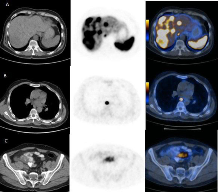
Results: Forty patients with a mean age of 48.1±15.80 years entered the study. Twenty-one (52.5%) were male and 19 (47.5%) female. In the studied patients, neuroendocrine tumor was present in 19 cases (47.5%) in pancreas and gastrointestinal tract, 9 (22.5%) in lung, 3 (7.5%) in mediastinum and adrenal gland, 6 cases (5%) in liver and 3 other sites. There was no significant association between mean age and gender with primary location of the tumor. The mean SUVmax was 11.62±20.02 and the the mean tumor size was 38.25±31.35 mm. The mean size of the metastasis was 40.55±24.53 mm. The mean percentage of ki-67 was 12.54±18.40. There was no significant correlation between SUVmax of the lesion and age (r=0.063, P=0.701), tumor size (r=-0.63, P=0.067) or Ki-67 (r=0.011, P=0.960). In 20 cases, metastases were reported, of which 14 were (70%) in the liver, 3 in the lungs (15%), 2 in the gastrointestinal and cervical lymph nodes, and 1 in the bones and pancreas(%5).
Conclusion: 68Ga-DOTA-peptide PET/CT could find the primary or metastasis sites of NETs with good quality images. In general, this modality can enhance the management in patients with NETs.


 分享
分享
 求助内容:
求助内容: 应助结果提醒方式:
 扫码关注我们
