Abdul Rahman Akkawi, Lynn Ezzeddine, Rita Chahinian, Firas Ershaid, Diala Merheb, Majd Mzeihem, Jean El-Cheikh, Mohamad Haidar
{"title":"Hepatic granuloma mimicking recurrent lymphoma on <sup>18</sup>F-FDG PET/CT in a patient with primary mediastinal diffuse large B-cell lymphoma.","authors":"Abdul Rahman Akkawi, Lynn Ezzeddine, Rita Chahinian, Firas Ershaid, Diala Merheb, Majd Mzeihem, Jean El-Cheikh, Mohamad Haidar","doi":"10.22038/AOJNMB.2021.56876.1396","DOIUrl":null,"url":null,"abstract":"<p><p><sup>18</sup>F-Flurodeoxyglucose (FDG) PET/CT has been considered the modality of choice in detecting, staging, restaging and following-up with lymphoma patients. However, it has an uncertain role in differentiating hepatic lymphomatous relapse from other granulomatous diseases such as in candidiasis or sarcoidosis. Therefore, it is important to correlate the imaging findings with other modalities such as ultrasound, CT scan, MRI, and histology to direct the diagnosis and treatment. We present a case of a 33-year-old woman with large B-cell lymphoma in complete remission following treatment presenting with neutropenic fever following her final cycle of chemotherapy. Ultrasound of the abdomen and enhanced CT scan of the abdomen and pelvis were negative. The FDG PET/CT scan showed multiple FDG-avid hypodense hepatic lesions that were suggestive either of lymphoproliferative involvement or nonmalignant process. However, MRI of the abdomen performed four days later was suggestive of an infectious process, rather than a lymphoproliferative disorder. A subsequent CT-guided biopsy of a hepatic lesion showed granulomatous inflammation, with no evidence of malignancy or Tuberculosis. The patient was started on Caspofungin followed by Fluconazole. After 5 weeks, the clinical condition resolved, and the subsequent FDG PET/CT showed complete resolution of the FDG-avid multiple hepatic lesions.</p>","PeriodicalId":8503,"journal":{"name":"Asia Oceania Journal of Nuclear Medicine and Biology","volume":"10 1","pages":"47-52"},"PeriodicalIF":0.0000,"publicationDate":"2022-01-01","publicationTypes":"Journal Article","fieldsOfStudy":null,"isOpenAccess":false,"openAccessPdf":"https://www.ncbi.nlm.nih.gov/pmc/articles/PMC8742851/pdf/","citationCount":"0","resultStr":null,"platform":"Semanticscholar","paperid":null,"PeriodicalName":"Asia Oceania Journal of Nuclear Medicine and Biology","FirstCategoryId":"1085","ListUrlMain":"https://doi.org/10.22038/AOJNMB.2021.56876.1396","RegionNum":0,"RegionCategory":null,"ArticlePicture":[],"TitleCN":null,"AbstractTextCN":null,"PMCID":null,"EPubDate":"","PubModel":"","JCR":"Q3","JCRName":"Medicine","Score":null,"Total":0}
引用次数: 0
Abstract
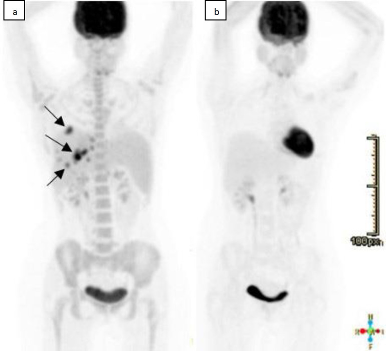
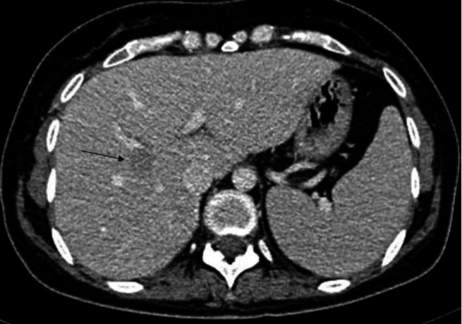
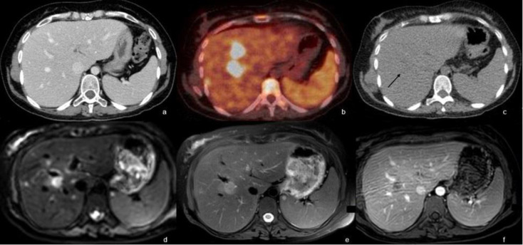
18F-Flurodeoxyglucose (FDG) PET/CT has been considered the modality of choice in detecting, staging, restaging and following-up with lymphoma patients. However, it has an uncertain role in differentiating hepatic lymphomatous relapse from other granulomatous diseases such as in candidiasis or sarcoidosis. Therefore, it is important to correlate the imaging findings with other modalities such as ultrasound, CT scan, MRI, and histology to direct the diagnosis and treatment. We present a case of a 33-year-old woman with large B-cell lymphoma in complete remission following treatment presenting with neutropenic fever following her final cycle of chemotherapy. Ultrasound of the abdomen and enhanced CT scan of the abdomen and pelvis were negative. The FDG PET/CT scan showed multiple FDG-avid hypodense hepatic lesions that were suggestive either of lymphoproliferative involvement or nonmalignant process. However, MRI of the abdomen performed four days later was suggestive of an infectious process, rather than a lymphoproliferative disorder. A subsequent CT-guided biopsy of a hepatic lesion showed granulomatous inflammation, with no evidence of malignancy or Tuberculosis. The patient was started on Caspofungin followed by Fluconazole. After 5 weeks, the clinical condition resolved, and the subsequent FDG PET/CT showed complete resolution of the FDG-avid multiple hepatic lesions.


 分享
分享
 求助内容:
求助内容: 应助结果提醒方式:
 扫码关注我们
