{"title":"Case of resected small-cell neuroendocrine carcinoma of the extrahepatic bile duct.","authors":"Hiroaki Sugita, Kazuya Maeda, Satoshi Nishikawa, Kenji Doden, Yasuo Hashizume","doi":"10.1093/jscr/rjac020","DOIUrl":null,"url":null,"abstract":"<p><p>Neuroendocrine carcinomas (NECs) arising from the extrahepatic bile duct (EHBD) are extremely rare, and their preoperative diagnosis is difficult. A small number of resected cases of EHBD NECs has been reported, and their prognosis is usually poor. A 62-year-old man presented with obstructive jaundice and liver disease. Radiological imaging revealed wall thickness and stricture of the distal common bile duct (CBD); however, lymph node or distant metastasis was not detected. Adenocarcinoma was detected on biopsy, and surgery was performed with a preoperative diagnosis of cholangiocarcinoma of the distal CBD. Pathological examination revealed adenocarcinoma of the CBD mucosa (20%) and NEC of the CBD wall (80%). The final pathological diagnosis was small-cell NEC of the EHBD. His post-operative course was good, and there was no recurrence for 4 months after surgery. Herein, we report a case of resected EHBD NEC and a literature review.</p>","PeriodicalId":47321,"journal":{"name":"Journal of Surgical Case Reports","volume":"2022 2","pages":"rjac020"},"PeriodicalIF":0.5000,"publicationDate":"2022-02-09","publicationTypes":"Journal Article","fieldsOfStudy":null,"isOpenAccess":false,"openAccessPdf":"https://www.ncbi.nlm.nih.gov/pmc/articles/PMC8828790/pdf/","citationCount":"0","resultStr":null,"platform":"Semanticscholar","paperid":null,"PeriodicalName":"Journal of Surgical Case Reports","FirstCategoryId":"1085","ListUrlMain":"https://doi.org/10.1093/jscr/rjac020","RegionNum":0,"RegionCategory":null,"ArticlePicture":[],"TitleCN":null,"AbstractTextCN":null,"PMCID":null,"EPubDate":"2022/2/1 0:00:00","PubModel":"eCollection","JCR":"Q4","JCRName":"SURGERY","Score":null,"Total":0}
引用次数: 0
Abstract
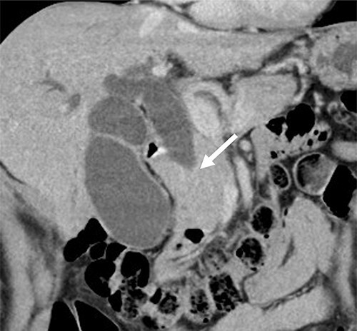
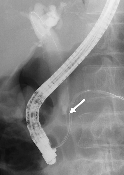
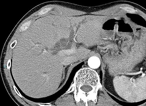
Neuroendocrine carcinomas (NECs) arising from the extrahepatic bile duct (EHBD) are extremely rare, and their preoperative diagnosis is difficult. A small number of resected cases of EHBD NECs has been reported, and their prognosis is usually poor. A 62-year-old man presented with obstructive jaundice and liver disease. Radiological imaging revealed wall thickness and stricture of the distal common bile duct (CBD); however, lymph node or distant metastasis was not detected. Adenocarcinoma was detected on biopsy, and surgery was performed with a preoperative diagnosis of cholangiocarcinoma of the distal CBD. Pathological examination revealed adenocarcinoma of the CBD mucosa (20%) and NEC of the CBD wall (80%). The final pathological diagnosis was small-cell NEC of the EHBD. His post-operative course was good, and there was no recurrence for 4 months after surgery. Herein, we report a case of resected EHBD NEC and a literature review.


 分享
分享
 求助内容:
求助内容: 应助结果提醒方式:
 扫码关注我们
