{"title":"How to stain nucleic acids and proteins in Miller spreads.","authors":"Lorena Zannino, Marco Biggiogera","doi":"10.4081/ejh.2022.3364","DOIUrl":null,"url":null,"abstract":"<p><p>The spread technique proposed by Miller and Beatty in 1969 allowed for the first time the visualization at transmission electron microscopy of nucleic acids and chromatin in an isolated and distended conformation. The final step of staining the spread chromatin is of critical importance because it can strongly influence the interpretation of the results. We evaluated different staining techniques and the most part of them provided a good result. Specifically, well contrasted micrographs were obtained when staining with H3PW12O40 (PTA), as originally proposed by Miller and Beatty, and with two alternatives proposed here: uranyl acetate or terbium citrate staining. Quite good contrast of the spread DNA could be achieved also by using Osmium Ammine; while no or few contrast of nucleic acids was observed by staining with KMnO₄ and H3PMo12O40 (PMA) respectively.</p>","PeriodicalId":50487,"journal":{"name":"European Journal of Histochemistry","volume":"66 1","pages":""},"PeriodicalIF":2.1000,"publicationDate":"2022-02-25","publicationTypes":"Journal Article","fieldsOfStudy":null,"isOpenAccess":false,"openAccessPdf":"https://ftp.ncbi.nlm.nih.gov/pub/pmc/oa_pdf/1e/a3/ejh-66-1-3364.PMC8883610.pdf","citationCount":"0","resultStr":null,"platform":"Semanticscholar","paperid":null,"PeriodicalName":"European Journal of Histochemistry","FirstCategoryId":"99","ListUrlMain":"https://doi.org/10.4081/ejh.2022.3364","RegionNum":4,"RegionCategory":"生物学","ArticlePicture":[],"TitleCN":null,"AbstractTextCN":null,"PMCID":null,"EPubDate":"","PubModel":"","JCR":"Q4","JCRName":"CELL BIOLOGY","Score":null,"Total":0}
引用次数: 0
Abstract
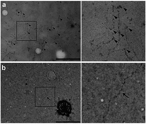
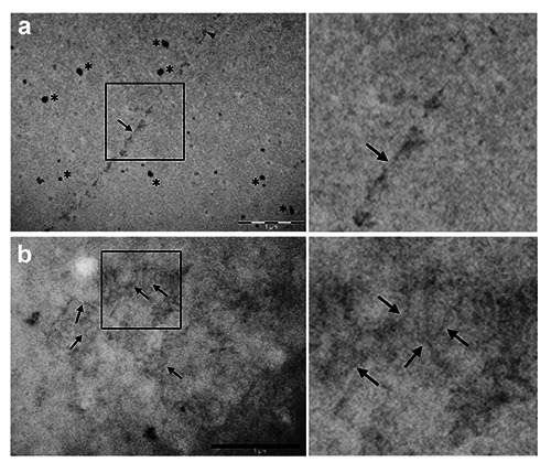
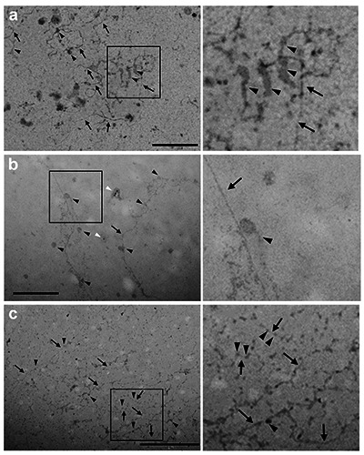
The spread technique proposed by Miller and Beatty in 1969 allowed for the first time the visualization at transmission electron microscopy of nucleic acids and chromatin in an isolated and distended conformation. The final step of staining the spread chromatin is of critical importance because it can strongly influence the interpretation of the results. We evaluated different staining techniques and the most part of them provided a good result. Specifically, well contrasted micrographs were obtained when staining with H3PW12O40 (PTA), as originally proposed by Miller and Beatty, and with two alternatives proposed here: uranyl acetate or terbium citrate staining. Quite good contrast of the spread DNA could be achieved also by using Osmium Ammine; while no or few contrast of nucleic acids was observed by staining with KMnO₄ and H3PMo12O40 (PMA) respectively.
期刊介绍:
The Journal publishes original papers concerning investigations by histochemical and immunohistochemical methods, and performed with the aid of light, super-resolution and electron microscopy, cytometry and imaging techniques. Coverage extends to:
functional cell and tissue biology in animals and plants;
cell differentiation and death;
cell-cell interaction and molecular trafficking;
biology of cell development and senescence;
nerve and muscle cell biology;
cellular basis of diseases.
The histochemical approach is nowadays essentially aimed at locating molecules in the very place where they exert their biological roles, and at describing dynamically specific chemical activities in living cells. Basic research on cell functional organization is essential for understanding the mechanisms underlying major biological processes such as differentiation, the control of tissue homeostasis, and the regulation of normal and tumor cell growth. Even more than in the past, the European Journal of Histochemistry, as a journal of functional cytology, represents the venue where cell scientists may present and discuss their original results, technical improvements and theories.


 分享
分享
 求助内容:
求助内容: 应助结果提醒方式:
 扫码关注我们
