{"title":"Diffuse Optical Tomography Using fNIRS Signals Measured from the Skull Surface of the Macaque Monkey.","authors":"Ryusuke Hayashi, Okito Yamashita, Toru Yamada, Hiroshi Kawaguchi, Noriyuki Higo","doi":"10.1093/texcom/tgab064","DOIUrl":null,"url":null,"abstract":"<p><p>Diffuse optical tomography (DOT), as a functional near-infrared spectroscopy (fNIRS) technique, can estimate three-dimensional (3D) images of the functional hemodynamic response in brain volume from measured optical signals. In this study, we applied DOT algorithms to the fNIRS data recorded from the surface of macaque monkeys' skulls when the animals performed food retrieval tasks using either the left- or right-hand under head-free conditions. The hemodynamic response images, reconstructed by DOT with a high sampling rate and fine voxel size, demonstrated significant activations at the upper limb regions of the primary motor area in the central sulcus and premotor, and parietal areas contralateral to the hands used in the tasks. The results were also reliable in terms of consistency across different recording dates. Time-series analyses of each brain area revealed preceding activity of premotor area to primary motor area consistent with previous physiological studies. Therefore, the fNIRS-DOT protocol demonstrated in this study provides reliable 3D functional brain images over a period of days under head-free conditions for region-of-interest-based time-series analysis.</p>","PeriodicalId":72551,"journal":{"name":"Cerebral cortex communications","volume":" ","pages":"tgab064"},"PeriodicalIF":0.0000,"publicationDate":"2021-11-10","publicationTypes":"Journal Article","fieldsOfStudy":null,"isOpenAccess":false,"openAccessPdf":"https://www.ncbi.nlm.nih.gov/pmc/articles/PMC8767783/pdf/","citationCount":"2","resultStr":null,"platform":"Semanticscholar","paperid":null,"PeriodicalName":"Cerebral cortex communications","FirstCategoryId":"1085","ListUrlMain":"https://doi.org/10.1093/texcom/tgab064","RegionNum":0,"RegionCategory":null,"ArticlePicture":[],"TitleCN":null,"AbstractTextCN":null,"PMCID":null,"EPubDate":"2022/1/1 0:00:00","PubModel":"eCollection","JCR":"","JCRName":"","Score":null,"Total":0}
引用次数: 2
Abstract
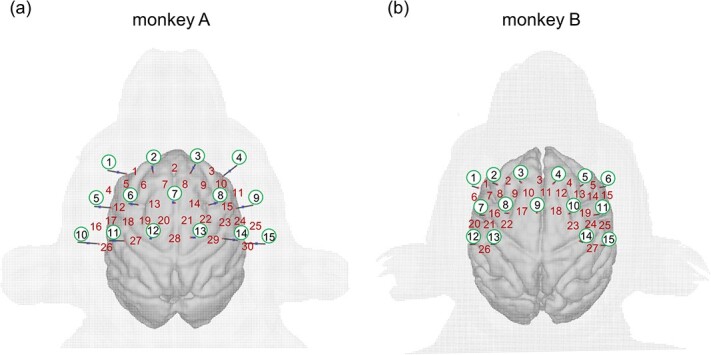
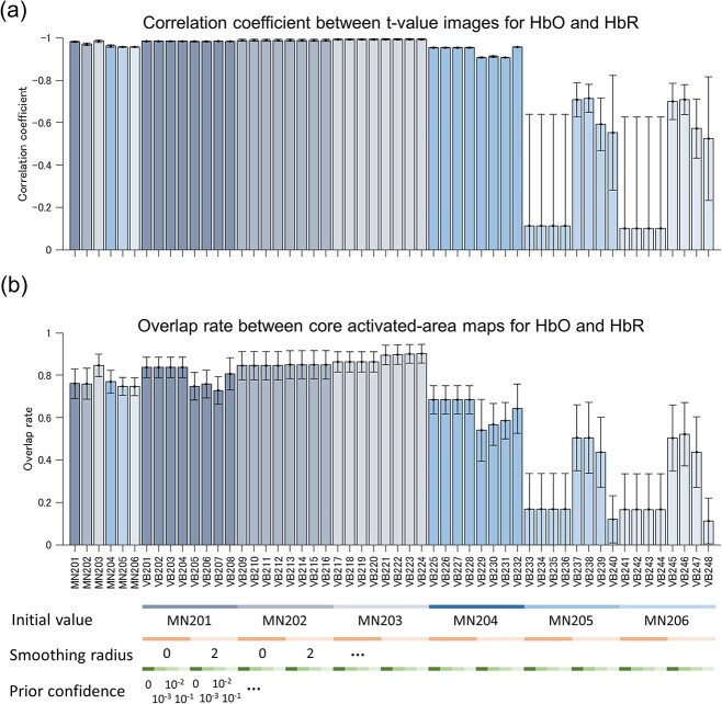
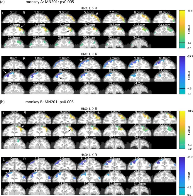
Diffuse optical tomography (DOT), as a functional near-infrared spectroscopy (fNIRS) technique, can estimate three-dimensional (3D) images of the functional hemodynamic response in brain volume from measured optical signals. In this study, we applied DOT algorithms to the fNIRS data recorded from the surface of macaque monkeys' skulls when the animals performed food retrieval tasks using either the left- or right-hand under head-free conditions. The hemodynamic response images, reconstructed by DOT with a high sampling rate and fine voxel size, demonstrated significant activations at the upper limb regions of the primary motor area in the central sulcus and premotor, and parietal areas contralateral to the hands used in the tasks. The results were also reliable in terms of consistency across different recording dates. Time-series analyses of each brain area revealed preceding activity of premotor area to primary motor area consistent with previous physiological studies. Therefore, the fNIRS-DOT protocol demonstrated in this study provides reliable 3D functional brain images over a period of days under head-free conditions for region-of-interest-based time-series analysis.


 分享
分享

 求助内容:
求助内容: 应助结果提醒方式:
 扫码关注我们
