First-time patellar dislocation with resultant habitual dislocation two years later, which was not demonstrated on plain X-rays halfway: a case report.
{"title":"First-time patellar dislocation with resultant habitual dislocation two years later, which was not demonstrated on plain X-rays halfway: a case report.","authors":"Satoshi Ohki, Hiroyuki Enomoto, Eiki Nomura, Hidenori Tanikawa, Yasuo Niki, Hideo Matsumoto, Yoshiaki Toyama, Yasunori Suda","doi":"10.1186/1758-2555-2-23","DOIUrl":null,"url":null,"abstract":"<p><p> We present an instructive case of habitual left patellar dislocation in which the patella had appeared odd due to lateral tilt relative to contralateral side, but had been radiologically confirmed to be on the trochlea at 1 year prior to the referral. An 11-year-old girl presented to our hospital 2 years after the left patella had dislocated with a 'giving way' when cutting to the left. Our physical and radiological examinations confirmed that the left patella was laterally tilted in the patellar groove with the knee in extension but was dislocated in flexion beyond 45°. In spite of these findings, she had been untreated at the previous hospital since all plain X-rays, including a skyline patellar view, had failed to demonstrate the dislocation. Consequently, in addition to reconstruction of medial patellofemoral ligament, she had to undergo a lateral retinacular release, which might have been unnecessary if treated earlier. This case illustrates that first-time patellar dislocation can gradually lead to habitual dislocation subsequently, and that cautious physical examinations in regard to patella tracking are essential since radiological examinations do not always reveal the pathophysiology of patellar instability.</p>","PeriodicalId":88316,"journal":{"name":"Sports medicine, arthroscopy, rehabilitation, therapy & technology : SMARTT","volume":" ","pages":"23"},"PeriodicalIF":0.0000,"publicationDate":"2010-09-14","publicationTypes":"Journal Article","fieldsOfStudy":null,"isOpenAccess":false,"openAccessPdf":"https://sci-hub-pdf.com/10.1186/1758-2555-2-23","citationCount":"1","resultStr":null,"platform":"Semanticscholar","paperid":null,"PeriodicalName":"Sports medicine, arthroscopy, rehabilitation, therapy & technology : SMARTT","FirstCategoryId":"1085","ListUrlMain":"https://doi.org/10.1186/1758-2555-2-23","RegionNum":0,"RegionCategory":null,"ArticlePicture":[],"TitleCN":null,"AbstractTextCN":null,"PMCID":null,"EPubDate":"","PubModel":"","JCR":"","JCRName":"","Score":null,"Total":0}
引用次数: 1
Abstract
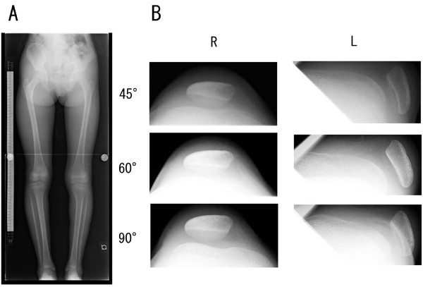
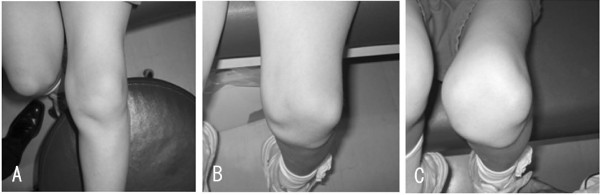
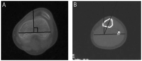
We present an instructive case of habitual left patellar dislocation in which the patella had appeared odd due to lateral tilt relative to contralateral side, but had been radiologically confirmed to be on the trochlea at 1 year prior to the referral. An 11-year-old girl presented to our hospital 2 years after the left patella had dislocated with a 'giving way' when cutting to the left. Our physical and radiological examinations confirmed that the left patella was laterally tilted in the patellar groove with the knee in extension but was dislocated in flexion beyond 45°. In spite of these findings, she had been untreated at the previous hospital since all plain X-rays, including a skyline patellar view, had failed to demonstrate the dislocation. Consequently, in addition to reconstruction of medial patellofemoral ligament, she had to undergo a lateral retinacular release, which might have been unnecessary if treated earlier. This case illustrates that first-time patellar dislocation can gradually lead to habitual dislocation subsequently, and that cautious physical examinations in regard to patella tracking are essential since radiological examinations do not always reveal the pathophysiology of patellar instability.


 分享
分享

 求助内容:
求助内容: 应助结果提醒方式:
 扫码关注我们
