Alessandro de Paula, Andrea Perdicchi, Augusto Pocobelli, Serena Fragiotta, Gianluca Scuderi
{"title":"The \"Topography\" of Glaucomatous Defect Using OCT and Visual Field Examination.","authors":"Alessandro de Paula, Andrea Perdicchi, Augusto Pocobelli, Serena Fragiotta, Gianluca Scuderi","doi":"10.5005/jp-journals-10078-1353","DOIUrl":null,"url":null,"abstract":"<p><strong>Aim: </strong>To describe the modifications in the superior and inferior retinal nerve fiber layer (RNFL) thickness regarding the distribution of the VF defects for the horizontal meridians in glaucomatous patients and the differences in the RNFL thickness topography between glaucomatous and healthy subjects.</p><p><strong>Methods: </strong>One hundred twenty eyes of 91 patients affected by glaucoma and 94 eyes of 51 normal patients were retrospectively reviewed. Computerized 30°VF (Octopus G1 Dynamic strategy) and optical coherence tomography (OCT) ONH and 3D disk analysis were performed in all cases. The RNFL thickness measures analyzed in both groups were superior-nasal (SN), superior-temporal (ST), inferior-nasal (IN), and inferior temporal (IT) sectors. The VFs were classified according to the distribution of the VF defect as for the horizontal meridian in the pattern deviation plot as superior, inferior, predominantly superior, or predominantly inferior.</p><p><strong>Result: </strong>In the glaucomatous group, 78 eyes (65%) showed a predominantly superior VF defect, while 38 eyes (32%) showed a predominantly inferior VF defect. Fifty-six eyes (46.7%) presented an exclusively superior, and 27/120 eyes (22.5%) presented an exclusively inferior VF defect. In the control group, the thickest RNFL sector was IT. The ST sector showed the thickest RNFL in presence of an exclusive superior VF defect. In case of an exclusive inferior VF defect, the thickest RNFL was the IT sector. VF showing superior defect presented a more altered MD than the VF with an inferior defect.</p><p><strong>Conclusion: </strong>Glaucomatous damage affects both the superior and inferior neural rim almost simultaneously. However, the neural rim loss seems to be asymmetric, involving the inferior or superior rim depending on the predominant involvement of the superior or inferior hemifield at the VF test. Particularly, the IT sector appears to be the most compromised in glaucomatous eyes. Therefore, the asymmetry between superior and inferior RNFL could support the diagnosis of glaucoma.</p><p><strong>How to cite this article: </strong>de Paula A, Perdicchi A, Pocobelli A, <i>et al.</i> The \"Topography\" of Glaucomatous Defect Using OCT and Visual Field Examination. J Curr Glaucoma Pract 2022;16(1):31-35.</p>","PeriodicalId":15419,"journal":{"name":"Journal of Current Glaucoma Practice","volume":" ","pages":"31-35"},"PeriodicalIF":0.0000,"publicationDate":"2022-01-01","publicationTypes":"Journal Article","fieldsOfStudy":null,"isOpenAccess":false,"openAccessPdf":"https://ftp.ncbi.nlm.nih.gov/pub/pmc/oa_pdf/ed/e6/jocgp-16-31.PMC9385384.pdf","citationCount":"0","resultStr":null,"platform":"Semanticscholar","paperid":null,"PeriodicalName":"Journal of Current Glaucoma Practice","FirstCategoryId":"1085","ListUrlMain":"https://doi.org/10.5005/jp-journals-10078-1353","RegionNum":0,"RegionCategory":null,"ArticlePicture":[],"TitleCN":null,"AbstractTextCN":null,"PMCID":null,"EPubDate":"","PubModel":"","JCR":"Q3","JCRName":"Medicine","Score":null,"Total":0}
引用次数: 0
Abstract
Aim: To describe the modifications in the superior and inferior retinal nerve fiber layer (RNFL) thickness regarding the distribution of the VF defects for the horizontal meridians in glaucomatous patients and the differences in the RNFL thickness topography between glaucomatous and healthy subjects.
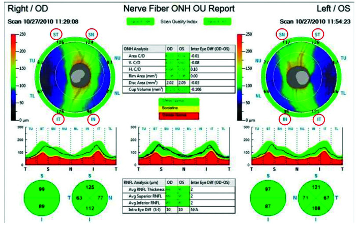
Methods: One hundred twenty eyes of 91 patients affected by glaucoma and 94 eyes of 51 normal patients were retrospectively reviewed. Computerized 30°VF (Octopus G1 Dynamic strategy) and optical coherence tomography (OCT) ONH and 3D disk analysis were performed in all cases. The RNFL thickness measures analyzed in both groups were superior-nasal (SN), superior-temporal (ST), inferior-nasal (IN), and inferior temporal (IT) sectors. The VFs were classified according to the distribution of the VF defect as for the horizontal meridian in the pattern deviation plot as superior, inferior, predominantly superior, or predominantly inferior.
Result: In the glaucomatous group, 78 eyes (65%) showed a predominantly superior VF defect, while 38 eyes (32%) showed a predominantly inferior VF defect. Fifty-six eyes (46.7%) presented an exclusively superior, and 27/120 eyes (22.5%) presented an exclusively inferior VF defect. In the control group, the thickest RNFL sector was IT. The ST sector showed the thickest RNFL in presence of an exclusive superior VF defect. In case of an exclusive inferior VF defect, the thickest RNFL was the IT sector. VF showing superior defect presented a more altered MD than the VF with an inferior defect.
Conclusion: Glaucomatous damage affects both the superior and inferior neural rim almost simultaneously. However, the neural rim loss seems to be asymmetric, involving the inferior or superior rim depending on the predominant involvement of the superior or inferior hemifield at the VF test. Particularly, the IT sector appears to be the most compromised in glaucomatous eyes. Therefore, the asymmetry between superior and inferior RNFL could support the diagnosis of glaucoma.
How to cite this article: de Paula A, Perdicchi A, Pocobelli A, et al. The "Topography" of Glaucomatous Defect Using OCT and Visual Field Examination. J Curr Glaucoma Pract 2022;16(1):31-35.
目的:探讨青光眼患者视网膜上、下神经纤维层厚度变化与水平经络VF缺损分布的关系,以及青光眼患者与正常人视网膜上、下神经纤维层厚度形貌的差异。方法:回顾性分析91例青光眼患者120只眼和51例正常患者94只眼的资料。所有病例均行计算机化30°VF (Octopus G1动态策略)、光学相干断层扫描(OCT) ONH和三维磁盘分析。两组的RNFL厚度测量分别为上鼻(SN)、上颞(ST)、下鼻(in)和下颞(IT)。根据VF缺陷在模式偏差图中水平子午线的分布情况,将VF分为上、下、主要上、主要下。结果:青光眼组78只眼(65%)表现为上视距缺损为主,38只眼(32%)表现为下视距缺损为主。56只眼(46.7%)表现为完全优越,27只眼(22.5%)表现为完全低下VF缺损。在对照组中,RNFL最厚的部分是IT。ST段显示最厚的RNFL存在排他的上VF缺陷。在排他性下位VF缺陷的情况下,最厚的RNFL是IT扇区。表现为上位缺损的VF比表现为下位缺损的VF表现出更大的MD改变。结论:青光眼损害几乎同时影响上、下神经圈。然而,神经边缘的丧失似乎是不对称的,根据VF测试中主要受累的是上半野还是下半野,神经边缘的受损是下或上边缘。特别是,在青光眼中,IT行业似乎是最容易受到损害的。因此,上、下RNFL的不对称性可以支持青光眼的诊断。如何引用本文:de Paula A, Perdicchi A, Pocobelli A等。利用OCT和视野检查青光眼缺损的“地形图”。中华青光眼杂志;2009;16(1):31-35。


 分享
分享
 求助内容:
求助内容: 应助结果提醒方式:
 扫码关注我们
