Sana Boudabbous, Marion Hamard, Essia Saiji, Karel Gorican, Pierre-Alexandre Poletti, Minerva Becker, Angeliki Neroladaki
{"title":"What morphological MRI features enable differentiation of low-grade from high-grade soft tissue sarcoma?","authors":"Sana Boudabbous, Marion Hamard, Essia Saiji, Karel Gorican, Pierre-Alexandre Poletti, Minerva Becker, Angeliki Neroladaki","doi":"10.1259/bjro.20210081","DOIUrl":null,"url":null,"abstract":"<p><strong>Objective: </strong>To assess the diagnostic performance of morphological MRI features separately and in combination for distinguishing low- from high-grade soft tissue sarcoma (STS).</p><p><strong>Methods and materials: </strong>We retrospectively analysed pre-treatment MRI examinations with T1, T2 with and without fat suppression (FS) and contrast-enhanced T1 obtained in 64 patients with STS categorized histologically as low (<i>n</i> = 21) versus high grade (<i>n</i> = 43). Two musculoskeletal radiologists blinded to histology evaluated MRI features. Diagnostic performance was calculated for each reader and for MRI features showing significant association with histology (<i>p</i> < 0.05). Logistic regression analysis was performed to develop a diagnostic model to identify high-grade STS.</p><p><strong>Results: </strong>Among all evaluated MRI features, only six features had adequate interobserver reproducibility (kappa>0.5). Multivariate logistic regression analysis revealed a significant association with tumour grade for lesion heterogeneity on FS images, intratumoural enhancement≥51% of tumour volume and peritumoural enhancement for both readers (<i>p</i> < 0.05). For both readers, the presence of each of the three features yielded odds ratios for high grade versus low grade from 4.4 to 9.1 (<i>p</i> < 0.05). The sum of the positive features for each reader independent of reader expertise yielded areas under the curve (AUCs) > 0.8. The presence of ≥2 positive features indicated a high risk for high-grade sarcoma, whereas ≤1 positive feature indicated a low-to-moderate risk.</p><p><strong>Conclusion: </strong>A diagnostic MRI score based on tumour heterogeneity, intratumoural and peritumoural enhancement enables identification of lesions that are likely to be high-grade as opposed to low-grade STS.</p><p><strong>Advances in knowledge: </strong>Tumour heterogeneity in Fat Suppression sequence, intratumoural and peritumoural enhancement is identified as signs of high-grade sarcoma.</p>","PeriodicalId":72419,"journal":{"name":"BJR open","volume":" ","pages":"20210081"},"PeriodicalIF":2.1000,"publicationDate":"2022-06-22","publicationTypes":"Journal Article","fieldsOfStudy":null,"isOpenAccess":false,"openAccessPdf":"https://www.ncbi.nlm.nih.gov/pmc/articles/PMC9459866/pdf/","citationCount":"4","resultStr":null,"platform":"Semanticscholar","paperid":null,"PeriodicalName":"BJR open","FirstCategoryId":"1085","ListUrlMain":"https://doi.org/10.1259/bjro.20210081","RegionNum":0,"RegionCategory":null,"ArticlePicture":[],"TitleCN":null,"AbstractTextCN":null,"PMCID":null,"EPubDate":"2022/1/1 0:00:00","PubModel":"eCollection","JCR":"","JCRName":"","Score":null,"Total":0}
引用次数: 4
Abstract
Objective: To assess the diagnostic performance of morphological MRI features separately and in combination for distinguishing low- from high-grade soft tissue sarcoma (STS).
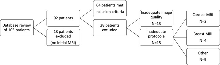
Methods and materials: We retrospectively analysed pre-treatment MRI examinations with T1, T2 with and without fat suppression (FS) and contrast-enhanced T1 obtained in 64 patients with STS categorized histologically as low (n = 21) versus high grade (n = 43). Two musculoskeletal radiologists blinded to histology evaluated MRI features. Diagnostic performance was calculated for each reader and for MRI features showing significant association with histology (p < 0.05). Logistic regression analysis was performed to develop a diagnostic model to identify high-grade STS.
Results: Among all evaluated MRI features, only six features had adequate interobserver reproducibility (kappa>0.5). Multivariate logistic regression analysis revealed a significant association with tumour grade for lesion heterogeneity on FS images, intratumoural enhancement≥51% of tumour volume and peritumoural enhancement for both readers (p < 0.05). For both readers, the presence of each of the three features yielded odds ratios for high grade versus low grade from 4.4 to 9.1 (p < 0.05). The sum of the positive features for each reader independent of reader expertise yielded areas under the curve (AUCs) > 0.8. The presence of ≥2 positive features indicated a high risk for high-grade sarcoma, whereas ≤1 positive feature indicated a low-to-moderate risk.
Conclusion: A diagnostic MRI score based on tumour heterogeneity, intratumoural and peritumoural enhancement enables identification of lesions that are likely to be high-grade as opposed to low-grade STS.
Advances in knowledge: Tumour heterogeneity in Fat Suppression sequence, intratumoural and peritumoural enhancement is identified as signs of high-grade sarcoma.


 分享
分享

 求助内容:
求助内容: 应助结果提醒方式:
 扫码关注我们
