{"title":"Cerebro-cerebellar interactions in nonhuman primates examined by optogenetic functional magnetic resonance imaging.","authors":"Naokazu Goda, Taku Hasegawa, Daisuke Koketsu, Satomi Chiken, Satomi Kikuta, Hiromi Sano, Kenta Kobayashi, Atsushi Nambu, Norihiro Sadato, Masaki Fukunaga","doi":"10.1093/texcom/tgac022","DOIUrl":null,"url":null,"abstract":"<p><p>Functional magnetic resonance imaging (fMRI) is a promising approach for the simultaneous and extensive scanning of whole-brain activities. Optogenetics is free from electrical and magnetic artifacts and is an ideal stimulation method for combined use with fMRI. However, the application of optogenetics in nonhuman primates (NHPs) remains limited. Recently, we developed an efficient optogenetic intracortical microstimulation method of the primary motor cortex (M1), which successfully induced forelimb movements in macaque monkeys. Here, we aimed to investigate how optogenetic M1 stimulation causes neural modulation in the local and remote brain regions in anesthetized monkeys using 7-tesla fMRI. We demonstrated that optogenetic stimulation of the M1 forelimb and hindlimb regions successfully evoked robust direct and remote fMRI activities. Prominent remote activities were detected in the anterior and posterior lobes in the contralateral cerebellum, which receive projections polysynaptically from the M1. We further demonstrated that the cerebro-cerebellar projections from these M1 regions were topographically organized, which is concordant with the somatotopic map in the cerebellar cortex previously reported in macaques and humans. The present study significantly enhances optogenetic fMRI in NHPs, resulting in profound understanding of the brain network, thereby accelerating the translation of findings from animal models to humans.</p>","PeriodicalId":72551,"journal":{"name":"Cerebral cortex communications","volume":" ","pages":"tgac022"},"PeriodicalIF":0.0000,"publicationDate":"2022-05-25","publicationTypes":"Journal Article","fieldsOfStudy":null,"isOpenAccess":false,"openAccessPdf":"https://www.ncbi.nlm.nih.gov/pmc/articles/PMC9233902/pdf/","citationCount":"0","resultStr":null,"platform":"Semanticscholar","paperid":null,"PeriodicalName":"Cerebral cortex communications","FirstCategoryId":"1085","ListUrlMain":"https://doi.org/10.1093/texcom/tgac022","RegionNum":0,"RegionCategory":null,"ArticlePicture":[],"TitleCN":null,"AbstractTextCN":null,"PMCID":null,"EPubDate":"2022/1/1 0:00:00","PubModel":"eCollection","JCR":"","JCRName":"","Score":null,"Total":0}
引用次数: 0
Abstract
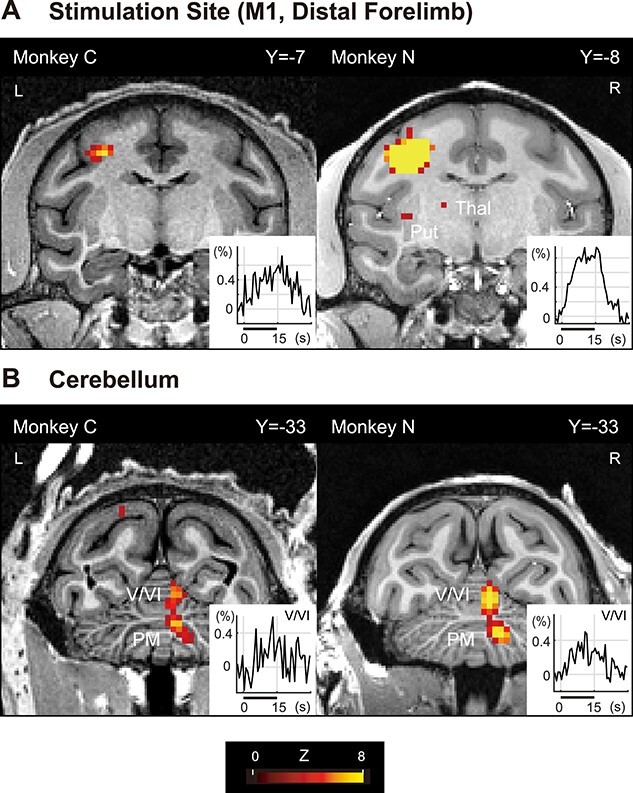
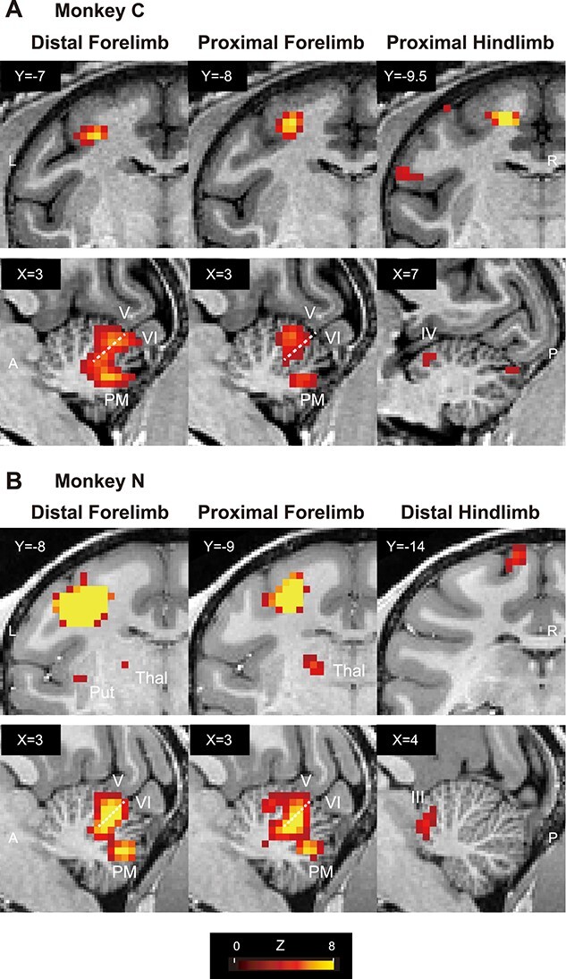
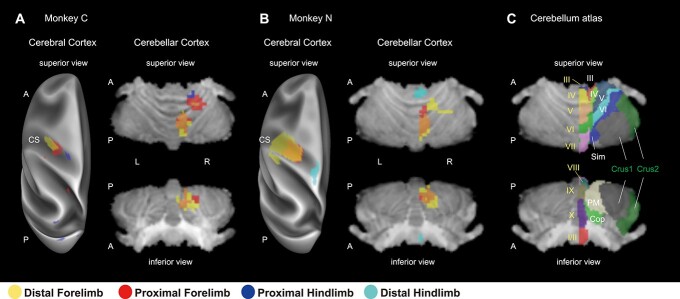
Functional magnetic resonance imaging (fMRI) is a promising approach for the simultaneous and extensive scanning of whole-brain activities. Optogenetics is free from electrical and magnetic artifacts and is an ideal stimulation method for combined use with fMRI. However, the application of optogenetics in nonhuman primates (NHPs) remains limited. Recently, we developed an efficient optogenetic intracortical microstimulation method of the primary motor cortex (M1), which successfully induced forelimb movements in macaque monkeys. Here, we aimed to investigate how optogenetic M1 stimulation causes neural modulation in the local and remote brain regions in anesthetized monkeys using 7-tesla fMRI. We demonstrated that optogenetic stimulation of the M1 forelimb and hindlimb regions successfully evoked robust direct and remote fMRI activities. Prominent remote activities were detected in the anterior and posterior lobes in the contralateral cerebellum, which receive projections polysynaptically from the M1. We further demonstrated that the cerebro-cerebellar projections from these M1 regions were topographically organized, which is concordant with the somatotopic map in the cerebellar cortex previously reported in macaques and humans. The present study significantly enhances optogenetic fMRI in NHPs, resulting in profound understanding of the brain network, thereby accelerating the translation of findings from animal models to humans.


 分享
分享

 求助内容:
求助内容: 应助结果提醒方式:
 扫码关注我们
