Mette Louise Gram Kjærulff, André H Dias, Peter Iversen, Lars Christian Gormsen, Karin Hjorthaug
{"title":"Early acquisition of [<sup>18</sup>F]FDOPA PET/CT imaging in patients with recurrent or residual medullary thyroid cancer is safe-and slightly better!","authors":"Mette Louise Gram Kjærulff, André H Dias, Peter Iversen, Lars Christian Gormsen, Karin Hjorthaug","doi":"10.1186/s41824-022-00140-7","DOIUrl":null,"url":null,"abstract":"<p><strong>Purpose: </strong>The aim of this study was to compare early (15 min) and late (60 min) [<sup>18</sup>F]FDOPA PET/CT acquisition times in the detection of recurrence/residual disease in medullary thyroid cancer (MTC) patients.</p><p><strong>Materials and methods: </strong>Thirty-two dual-phase [<sup>18</sup>F]FDOPA PET scans were retrospectively reviewed. Scan indications were (1) suspected recurrence of MTC, (2) treatment monitoring, or (3) restaging. In four scans, no final verification could be obtained, and one scan was excluded due to non-consistency with the acquisition protocol. Images were analyzed visually and semiquantitatively (using SUV<sub>max</sub>). On both per-scan and per-lesion basis, early (median time 15 min) and late (median time 60 min) acquisition were compared by number and SUV<sub>max</sub> of detected MTC lesions, and a washout rate between the two acquisitions was calculated. Sensitivity and specificity of early and late acquisition were also compared.</p><p><strong>Results: </strong>Out of the 27 eligible PET scans, twenty were classified as PET positive and 7 as PET negative. By subsequent histology and/or combination of imaging and clinical data during follow-up, the MTC diagnosis was verified, showing a scan-based sensitivity and specificity of 100% and 87.5%, respectively, for the early acquisition, and for the late acquisition both were 100%. However, there were no statistically significant difference in detection rate between the two acquisitions. Lesions on the early acquisition were significantly more intense compared to lesions on the late acquisition (median washout rate of - 33% (- 57 to + 50%)).</p><p><strong>Conclusion: </strong>Our study confirms that it is safe to omit the late [<sup>18</sup>F]FDOPA PET/CT acquisition in the detection of recurrent/residual MTC.</p>","PeriodicalId":36160,"journal":{"name":"European Journal of Hybrid Imaging","volume":" ","pages":"20"},"PeriodicalIF":1.2000,"publicationDate":"2022-08-25","publicationTypes":"Journal Article","fieldsOfStudy":null,"isOpenAccess":false,"openAccessPdf":"https://www.ncbi.nlm.nih.gov/pmc/articles/PMC9402850/pdf/","citationCount":"1","resultStr":null,"platform":"Semanticscholar","paperid":null,"PeriodicalName":"European Journal of Hybrid Imaging","FirstCategoryId":"1085","ListUrlMain":"https://doi.org/10.1186/s41824-022-00140-7","RegionNum":0,"RegionCategory":null,"ArticlePicture":[],"TitleCN":null,"AbstractTextCN":null,"PMCID":null,"EPubDate":"","PubModel":"","JCR":"Q3","JCRName":"RADIOLOGY, NUCLEAR MEDICINE & MEDICAL IMAGING","Score":null,"Total":0}
引用次数: 1
Abstract
Purpose: The aim of this study was to compare early (15 min) and late (60 min) [18F]FDOPA PET/CT acquisition times in the detection of recurrence/residual disease in medullary thyroid cancer (MTC) patients.
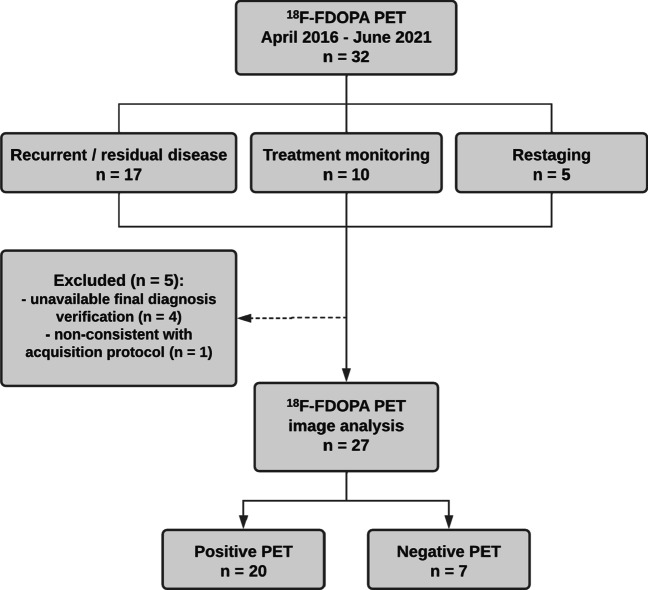
Materials and methods: Thirty-two dual-phase [18F]FDOPA PET scans were retrospectively reviewed. Scan indications were (1) suspected recurrence of MTC, (2) treatment monitoring, or (3) restaging. In four scans, no final verification could be obtained, and one scan was excluded due to non-consistency with the acquisition protocol. Images were analyzed visually and semiquantitatively (using SUVmax). On both per-scan and per-lesion basis, early (median time 15 min) and late (median time 60 min) acquisition were compared by number and SUVmax of detected MTC lesions, and a washout rate between the two acquisitions was calculated. Sensitivity and specificity of early and late acquisition were also compared.
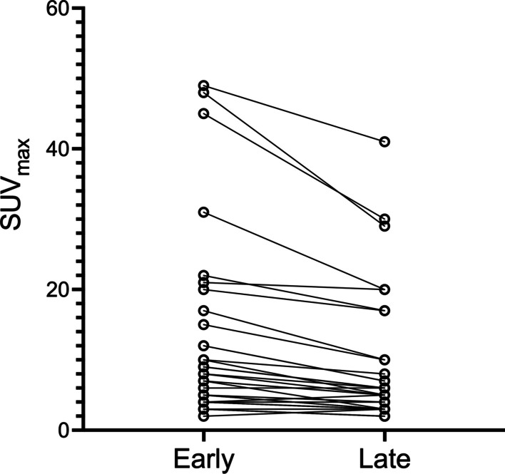
Results: Out of the 27 eligible PET scans, twenty were classified as PET positive and 7 as PET negative. By subsequent histology and/or combination of imaging and clinical data during follow-up, the MTC diagnosis was verified, showing a scan-based sensitivity and specificity of 100% and 87.5%, respectively, for the early acquisition, and for the late acquisition both were 100%. However, there were no statistically significant difference in detection rate between the two acquisitions. Lesions on the early acquisition were significantly more intense compared to lesions on the late acquisition (median washout rate of - 33% (- 57 to + 50%)).
Conclusion: Our study confirms that it is safe to omit the late [18F]FDOPA PET/CT acquisition in the detection of recurrent/residual MTC.


 分享
分享
 求助内容:
求助内容: 应助结果提醒方式:
 扫码关注我们
