Comparison of Diffusion Tensor Imaging Metrics in Normal-Appearing White Matter to Cerebrovascular Lesions and Correlation with Cerebrovascular Disease Risk Factors and Severity.
Seyyed M H Haddad, Christopher J M Scott, Miracle Ozzoude, Courtney Berezuk, Melissa Holmes, Sabrina Adamo, Joel Ramirez, Stephen R Arnott, Nuwan D Nanayakkara, Malcolm Binns, Derek Beaton, Wendy Lou, Kelly Sunderland, Sujeevini Sujanthan, Jane Lawrence, Donna Kwan, Brian Tan, Leanne Casaubon, Jennifer Mandzia, Demetrios Sahlas, Gustavo Saposnik, Ayman Hassan, Brian Levine, Paula McLaughlin, J B Orange, Angela Roberts, Angela Troyer, Sandra E Black, Dar Dowlatshahi, Stephen C Strother, Richard H Swartz, Sean Symons, Manuel Montero-Odasso, Ondri Investigators, Robert Bartha
{"title":"Comparison of Diffusion Tensor Imaging Metrics in Normal-Appearing White Matter to Cerebrovascular Lesions and Correlation with Cerebrovascular Disease Risk Factors and Severity.","authors":"Seyyed M H Haddad, Christopher J M Scott, Miracle Ozzoude, Courtney Berezuk, Melissa Holmes, Sabrina Adamo, Joel Ramirez, Stephen R Arnott, Nuwan D Nanayakkara, Malcolm Binns, Derek Beaton, Wendy Lou, Kelly Sunderland, Sujeevini Sujanthan, Jane Lawrence, Donna Kwan, Brian Tan, Leanne Casaubon, Jennifer Mandzia, Demetrios Sahlas, Gustavo Saposnik, Ayman Hassan, Brian Levine, Paula McLaughlin, J B Orange, Angela Roberts, Angela Troyer, Sandra E Black, Dar Dowlatshahi, Stephen C Strother, Richard H Swartz, Sean Symons, Manuel Montero-Odasso, Ondri Investigators, Robert Bartha","doi":"10.1155/2022/5860364","DOIUrl":null,"url":null,"abstract":"<p><p>Alterations in tissue microstructure in normal-appearing white matter (NAWM), specifically measured by diffusion tensor imaging (DTI) fractional anisotropy (FA), have been associated with cognitive outcomes following stroke. The purpose of this study was to comprehensively compare conventional DTI measures of tissue microstructure in NAWM to diverse vascular brain lesions in people with cerebrovascular disease (CVD) and to examine associations between FA in NAWM and cerebrovascular risk factors. DTI metrics including fractional anisotropy (FA), mean diffusivity (MD), axial diffusivity (AD), and radial diffusivity (RD) were measured in cerebral tissues and cerebrovascular anomalies from 152 people with CVD participating in the Ontario Neurodegenerative Disease Research Initiative (ONDRI). Ten cerebral tissue types were segmented including NAWM, and vascular lesions including stroke, periventricular and deep white matter hyperintensities, periventricular and deep lacunar infarcts, and perivascular spaces (PVS) using T<sub>1</sub>-weighted, proton density-weighted, T<sub>2</sub>-weighted, and fluid attenuated inversion recovery MRI scans. Mean DTI metrics were measured in each tissue region using a previously developed DTI processing pipeline and compared between tissues using multivariate analysis of covariance. Associations between FA in NAWM and several CVD risk factors were also examined. DTI metrics in vascular lesions differed significantly from healthy tissue. Specifically, all tissue types had significantly different MD values, while FA was also found to be different in most tissue types. FA in NAWM was inversely related to hypertension and modified Rankin scale (mRS). This study demonstrated the differences between conventional DTI metrics, FA, MD, AD, and RD, in cerebral vascular lesions and healthy tissue types. Therefore, incorporating DTI to characterize the integrity of the tissue microstructure could help to define the extent and severity of various brain vascular anomalies. The association between FA within NAWM and clinical evaluation of hypertension and disability provides further evidence that white matter microstructural integrity is impacted by cerebrovascular function.</p>","PeriodicalId":47063,"journal":{"name":"International Journal of Biomedical Imaging","volume":" ","pages":"5860364"},"PeriodicalIF":1.3000,"publicationDate":"2022-10-21","publicationTypes":"Journal Article","fieldsOfStudy":null,"isOpenAccess":false,"openAccessPdf":"https://www.ncbi.nlm.nih.gov/pmc/articles/PMC9616672/pdf/","citationCount":"2","resultStr":null,"platform":"Semanticscholar","paperid":null,"PeriodicalName":"International Journal of Biomedical Imaging","FirstCategoryId":"1085","ListUrlMain":"https://doi.org/10.1155/2022/5860364","RegionNum":0,"RegionCategory":null,"ArticlePicture":[],"TitleCN":null,"AbstractTextCN":null,"PMCID":null,"EPubDate":"2022/1/1 0:00:00","PubModel":"eCollection","JCR":"Q2","JCRName":"ENGINEERING, BIOMEDICAL","Score":null,"Total":0}
引用次数: 2
Abstract
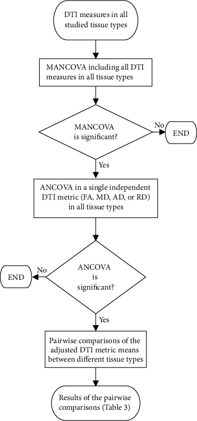
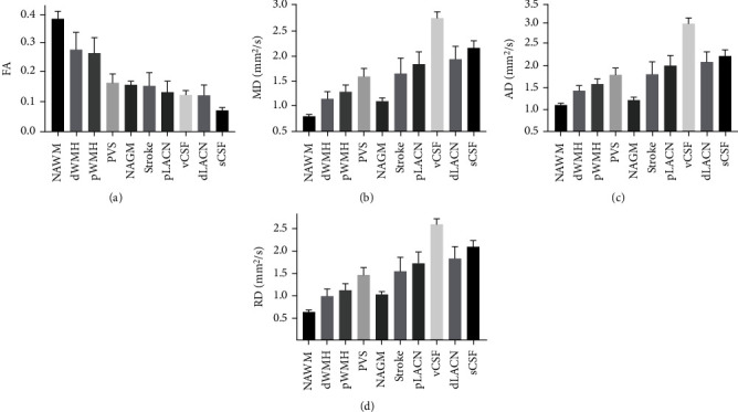
Alterations in tissue microstructure in normal-appearing white matter (NAWM), specifically measured by diffusion tensor imaging (DTI) fractional anisotropy (FA), have been associated with cognitive outcomes following stroke. The purpose of this study was to comprehensively compare conventional DTI measures of tissue microstructure in NAWM to diverse vascular brain lesions in people with cerebrovascular disease (CVD) and to examine associations between FA in NAWM and cerebrovascular risk factors. DTI metrics including fractional anisotropy (FA), mean diffusivity (MD), axial diffusivity (AD), and radial diffusivity (RD) were measured in cerebral tissues and cerebrovascular anomalies from 152 people with CVD participating in the Ontario Neurodegenerative Disease Research Initiative (ONDRI). Ten cerebral tissue types were segmented including NAWM, and vascular lesions including stroke, periventricular and deep white matter hyperintensities, periventricular and deep lacunar infarcts, and perivascular spaces (PVS) using T1-weighted, proton density-weighted, T2-weighted, and fluid attenuated inversion recovery MRI scans. Mean DTI metrics were measured in each tissue region using a previously developed DTI processing pipeline and compared between tissues using multivariate analysis of covariance. Associations between FA in NAWM and several CVD risk factors were also examined. DTI metrics in vascular lesions differed significantly from healthy tissue. Specifically, all tissue types had significantly different MD values, while FA was also found to be different in most tissue types. FA in NAWM was inversely related to hypertension and modified Rankin scale (mRS). This study demonstrated the differences between conventional DTI metrics, FA, MD, AD, and RD, in cerebral vascular lesions and healthy tissue types. Therefore, incorporating DTI to characterize the integrity of the tissue microstructure could help to define the extent and severity of various brain vascular anomalies. The association between FA within NAWM and clinical evaluation of hypertension and disability provides further evidence that white matter microstructural integrity is impacted by cerebrovascular function.
期刊介绍:
The International Journal of Biomedical Imaging is managed by a board of editors comprising internationally renowned active researchers. The journal is freely accessible online and also offered for purchase in print format. It employs a web-based review system to ensure swift turnaround times while maintaining high standards. In addition to regular issues, special issues are organized by guest editors. The subject areas covered include (but are not limited to):
Digital radiography and tomosynthesis
X-ray computed tomography (CT)
Magnetic resonance imaging (MRI)
Single photon emission computed tomography (SPECT)
Positron emission tomography (PET)
Ultrasound imaging
Diffuse optical tomography, coherence, fluorescence, bioluminescence tomography, impedance tomography
Neutron imaging for biomedical applications
Magnetic and optical spectroscopy, and optical biopsy
Optical, electron, scanning tunneling/atomic force microscopy
Small animal imaging
Functional, cellular, and molecular imaging
Imaging assays for screening and molecular analysis
Microarray image analysis and bioinformatics
Emerging biomedical imaging techniques
Imaging modality fusion
Biomedical imaging instrumentation
Biomedical image processing, pattern recognition, and analysis
Biomedical image visualization, compression, transmission, and storage
Imaging and modeling related to systems biology and systems biomedicine
Applied mathematics, applied physics, and chemistry related to biomedical imaging
Grid-enabling technology for biomedical imaging and informatics


 分享
分享
 求助内容:
求助内容: 应助结果提醒方式:
 扫码关注我们
