{"title":"Solitary Leptomeningeal Metastasis from Lung Cancer: A Case Report.","authors":"Keita Yokawa, Yuji Matsumoto, Keina Nagakita, Yoko Shinno, Kenichiro Kudo, Nanami Niguma, Kosaku Suenobu, Hideyuki Yoshida","doi":"10.2176/jns-nmc.2022-0113","DOIUrl":null,"url":null,"abstract":"<p><p>Leptomeningeal metastasis (LM) is a rare but devastating cancer complication. LM occurs when cancer spreads into the leptomeningeal layer or cerebrospinal fluid. Intracranial magnetic resonance (MR) images of LM are characterized by the diffuse enhancement of the leptomeninges along the cerebral sulci, cerebellar folia, and cranial nerves. Here, we report an extremely rare case of LM with an atypical MR image revealing tumor mass confinement to the arachnoid membrane. The case involves an 85-year-old man who was referred to our hospital with a three-day history of dysarthria. Radiological examination revealed a solid lesion with heterogeneous enhancement and a cystic component in the extra-axial region of the right parietal lobe. Upon subsequent general examination, multiple lung cancer metastases were suspected. The patient underwent gross total resection of the brain mass in the right parietal region. Although the tumor slightly adhered to the dura mater, it was sharply demarcated from the surrounding parenchyma and pia mater. Based on pathological examination, the tumor was diagnosed as small cell lung cancer metastasis. This metastatic brain tumor was exclusively confined to the arachnoid membrane and, except for a few blood vessels, the dura mater was not infiltrated by metastatic tumor cells. To our knowledge, this is the first reported case of LM in which the tumor mass is confined only to the arachnoid membrane. Thus, in cases with atypical MR images, a general examination considering the possibility of LM is important for prompt and accurate diagnosis.</p>","PeriodicalId":19260,"journal":{"name":"NMC Case Report Journal","volume":" ","pages":"323-328"},"PeriodicalIF":0.0000,"publicationDate":"2022-09-23","publicationTypes":"Journal Article","fieldsOfStudy":null,"isOpenAccess":false,"openAccessPdf":"https://ftp.ncbi.nlm.nih.gov/pub/pmc/oa_pdf/30/68/2188-4226-9-0323.PMC9560545.pdf","citationCount":"1","resultStr":null,"platform":"Semanticscholar","paperid":null,"PeriodicalName":"NMC Case Report Journal","FirstCategoryId":"1085","ListUrlMain":"https://doi.org/10.2176/jns-nmc.2022-0113","RegionNum":0,"RegionCategory":null,"ArticlePicture":[],"TitleCN":null,"AbstractTextCN":null,"PMCID":null,"EPubDate":"2022/1/1 0:00:00","PubModel":"eCollection","JCR":"","JCRName":"","Score":null,"Total":0}
引用次数: 1
Abstract
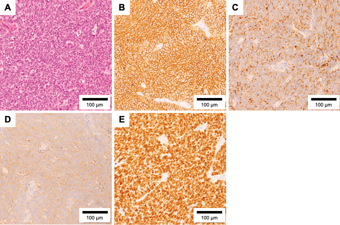
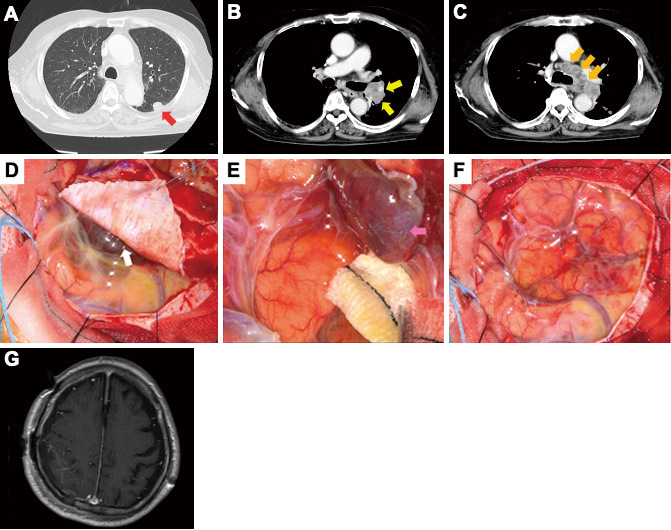
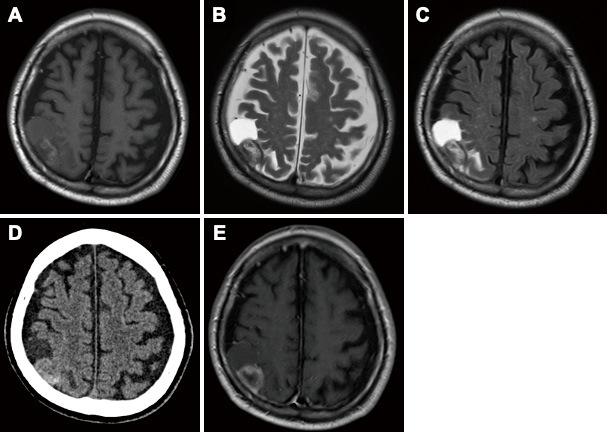
Leptomeningeal metastasis (LM) is a rare but devastating cancer complication. LM occurs when cancer spreads into the leptomeningeal layer or cerebrospinal fluid. Intracranial magnetic resonance (MR) images of LM are characterized by the diffuse enhancement of the leptomeninges along the cerebral sulci, cerebellar folia, and cranial nerves. Here, we report an extremely rare case of LM with an atypical MR image revealing tumor mass confinement to the arachnoid membrane. The case involves an 85-year-old man who was referred to our hospital with a three-day history of dysarthria. Radiological examination revealed a solid lesion with heterogeneous enhancement and a cystic component in the extra-axial region of the right parietal lobe. Upon subsequent general examination, multiple lung cancer metastases were suspected. The patient underwent gross total resection of the brain mass in the right parietal region. Although the tumor slightly adhered to the dura mater, it was sharply demarcated from the surrounding parenchyma and pia mater. Based on pathological examination, the tumor was diagnosed as small cell lung cancer metastasis. This metastatic brain tumor was exclusively confined to the arachnoid membrane and, except for a few blood vessels, the dura mater was not infiltrated by metastatic tumor cells. To our knowledge, this is the first reported case of LM in which the tumor mass is confined only to the arachnoid membrane. Thus, in cases with atypical MR images, a general examination considering the possibility of LM is important for prompt and accurate diagnosis.


 分享
分享

 求助内容:
求助内容: 应助结果提醒方式:
 扫码关注我们
