Structural plasticity of motor cortices assessed by voxel-based morphometry and immunohistochemical analysis following internal capsular infarcts in macaque monkeys.
{"title":"Structural plasticity of motor cortices assessed by voxel-based morphometry and immunohistochemical analysis following internal capsular infarcts in macaque monkeys.","authors":"Kohei Matsuda, Kazuaki Nagasaka, Junpei Kato, Ichiro Takashima, Noriyuki Higo","doi":"10.1093/texcom/tgac046","DOIUrl":null,"url":null,"abstract":"<p><p>Compensatory plastic changes in the remaining intact brain regions are supposedly involved in functional recovery following stroke. Previously, a compensatory increase in cortical activation occurred in the ventral premotor cortex (PMv), which contributed to the recovery of dexterous hand movement in a macaque model of unilateral internal capsular infarcts. Herein, we investigated the structural plastic changes underlying functional changes together with voxel-based morphometry (VBM) analysis of magnetic resonance imaging data and immunohistochemical analysis using SMI-32 antibody in a macaque model. Unilateral internal capsular infarcts were pharmacologically induced in 5 macaques, and another 5 macaques were used as intact controls for immunohistochemical analysis. Three months post infarcts, we observed significant increases in the gray matter volume (GMV) and the dendritic arborization of layer V pyramidal neurons in the contralesional rostral PMv (F5) as well as the primary motor cortex (M1). The histological analysis revealed shrinkage of neuronal soma and dendrites in the ipsilesional M1 and several premotor cortices, despite not always detecting GMV reduction by VBM analysis. In conclusion, compensatory structural changes occur in the contralesional F5 and M1 during motor recovery following internal capsular infarcts, and the dendritic growth of pyramidal neurons is partially correlated with GMV increase.</p>","PeriodicalId":72551,"journal":{"name":"Cerebral cortex communications","volume":" ","pages":"tgac046"},"PeriodicalIF":0.0000,"publicationDate":"2022-11-08","publicationTypes":"Journal Article","fieldsOfStudy":null,"isOpenAccess":false,"openAccessPdf":"https://www.ncbi.nlm.nih.gov/pmc/articles/PMC9706438/pdf/","citationCount":"0","resultStr":null,"platform":"Semanticscholar","paperid":null,"PeriodicalName":"Cerebral cortex communications","FirstCategoryId":"1085","ListUrlMain":"https://doi.org/10.1093/texcom/tgac046","RegionNum":0,"RegionCategory":null,"ArticlePicture":[],"TitleCN":null,"AbstractTextCN":null,"PMCID":null,"EPubDate":"2022/1/1 0:00:00","PubModel":"eCollection","JCR":"","JCRName":"","Score":null,"Total":0}
引用次数: 0
Abstract
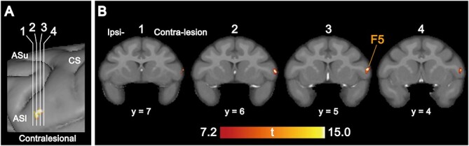
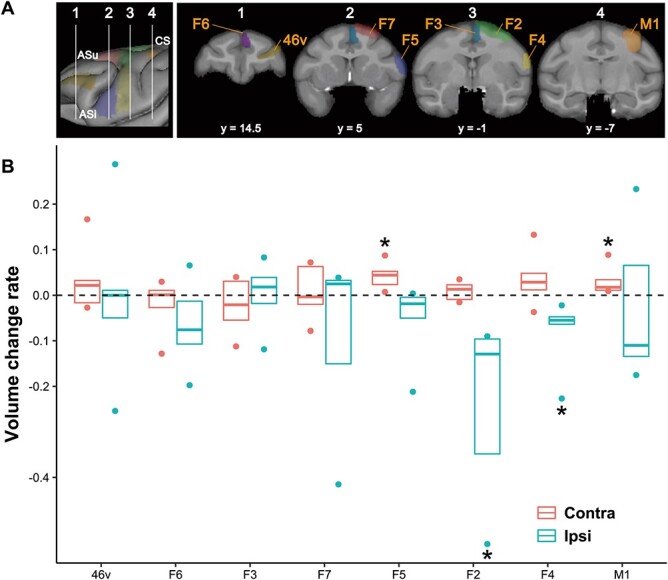
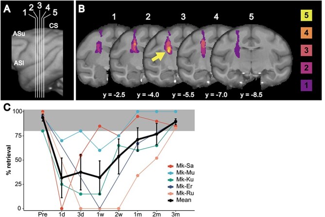
Compensatory plastic changes in the remaining intact brain regions are supposedly involved in functional recovery following stroke. Previously, a compensatory increase in cortical activation occurred in the ventral premotor cortex (PMv), which contributed to the recovery of dexterous hand movement in a macaque model of unilateral internal capsular infarcts. Herein, we investigated the structural plastic changes underlying functional changes together with voxel-based morphometry (VBM) analysis of magnetic resonance imaging data and immunohistochemical analysis using SMI-32 antibody in a macaque model. Unilateral internal capsular infarcts were pharmacologically induced in 5 macaques, and another 5 macaques were used as intact controls for immunohistochemical analysis. Three months post infarcts, we observed significant increases in the gray matter volume (GMV) and the dendritic arborization of layer V pyramidal neurons in the contralesional rostral PMv (F5) as well as the primary motor cortex (M1). The histological analysis revealed shrinkage of neuronal soma and dendrites in the ipsilesional M1 and several premotor cortices, despite not always detecting GMV reduction by VBM analysis. In conclusion, compensatory structural changes occur in the contralesional F5 and M1 during motor recovery following internal capsular infarcts, and the dendritic growth of pyramidal neurons is partially correlated with GMV increase.


 分享
分享

 求助内容:
求助内容: 应助结果提醒方式:
 扫码关注我们
