Murat Kasıkcı, Aylin Karalezli, Özgür Eroğul, Hamidu Hamisi Gobeka
{"title":"Quantifying contact lens-related changes in keratoconus corneal topographic indices: an updated Pentacam Scheimpflug imaging analysis.","authors":"Murat Kasıkcı, Aylin Karalezli, Özgür Eroğul, Hamidu Hamisi Gobeka","doi":"10.22336/rjo.2022.47","DOIUrl":null,"url":null,"abstract":"<p><p><b>Purpose:</b> Slowing ectasia progression is critical for maintaining visual potential in keratoconus (KC), for which various therapeutic approaches have been implemented. A Pentacam Scheimpflug imaging device was used to quantify contact lens (CL)-related changes in keratoconus corneal topographic indices. <b>Methods:</b> Thirty KC patients (group 1; 60 eyes) were using one of the three CL (rigid gas-permeable CL (RGPCL)-10, hybrid CL (HCL)-10, and scleral CL (SCL)-10 patients). A control group included 30 KC patients (group 2; 60 eyes) not using CLs due to intolerance or inappropriateness. The Pentacam® HR Scheimpflug imaging device was used to measure topographic indices such as Km anterior, Km posterior, K max, corneal thickness (CT, corneal central, apex, and thinnest), corneal volume (CV), anterior chamber volume (ACV), and anterior chamber depth (ACD) at baseline, 3rd, 6th, and 12th months. <b>Results:</b> The mean ages for groups 1 and 2 were 32±10 and 31±09 years, respectively. Group 1 had a lower but statistically significant change in K max than group 2 (<i>p</i><0.038). Also, group 1 had a minor but non-significant decrease in anterior and posterior keratometry values compared to group 2 (pKm ant. right/ left eye = 0.063/ 0.065 and 0.087/ 0.094, respectively). RGPCL users had significant changes in central CT, thinnest CT and ACD (<i>p</i><0.041). SCL users had more stable changes than other CLs for the thinnest CT along with significant changes in K max, pachy apex and ACV (<i>p</i><0.036). HCL users had significantly higher K max stability (<i>p</i><0.039). <b>Conclusion:</b> Regular use of appropriate therapeutic CLs may help to stabilize corneal deformity, thereby slowing changes in corneal topographic indices in KC.</p>","PeriodicalId":21385,"journal":{"name":"Romanian journal of ophthalmology","volume":"66 3","pages":"245-256"},"PeriodicalIF":0.0000,"publicationDate":"2022-07-01","publicationTypes":"Journal Article","fieldsOfStudy":null,"isOpenAccess":false,"openAccessPdf":"https://www.ncbi.nlm.nih.gov/pmc/articles/PMC9585500/pdf/","citationCount":"0","resultStr":null,"platform":"Semanticscholar","paperid":null,"PeriodicalName":"Romanian journal of ophthalmology","FirstCategoryId":"1085","ListUrlMain":"https://doi.org/10.22336/rjo.2022.47","RegionNum":0,"RegionCategory":null,"ArticlePicture":[],"TitleCN":null,"AbstractTextCN":null,"PMCID":null,"EPubDate":"","PubModel":"","JCR":"","JCRName":"","Score":null,"Total":0}
引用次数: 0
Abstract
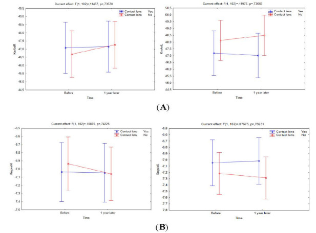
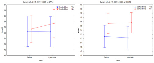
Purpose: Slowing ectasia progression is critical for maintaining visual potential in keratoconus (KC), for which various therapeutic approaches have been implemented. A Pentacam Scheimpflug imaging device was used to quantify contact lens (CL)-related changes in keratoconus corneal topographic indices. Methods: Thirty KC patients (group 1; 60 eyes) were using one of the three CL (rigid gas-permeable CL (RGPCL)-10, hybrid CL (HCL)-10, and scleral CL (SCL)-10 patients). A control group included 30 KC patients (group 2; 60 eyes) not using CLs due to intolerance or inappropriateness. The Pentacam® HR Scheimpflug imaging device was used to measure topographic indices such as Km anterior, Km posterior, K max, corneal thickness (CT, corneal central, apex, and thinnest), corneal volume (CV), anterior chamber volume (ACV), and anterior chamber depth (ACD) at baseline, 3rd, 6th, and 12th months. Results: The mean ages for groups 1 and 2 were 32±10 and 31±09 years, respectively. Group 1 had a lower but statistically significant change in K max than group 2 (p<0.038). Also, group 1 had a minor but non-significant decrease in anterior and posterior keratometry values compared to group 2 (pKm ant. right/ left eye = 0.063/ 0.065 and 0.087/ 0.094, respectively). RGPCL users had significant changes in central CT, thinnest CT and ACD (p<0.041). SCL users had more stable changes than other CLs for the thinnest CT along with significant changes in K max, pachy apex and ACV (p<0.036). HCL users had significantly higher K max stability (p<0.039). Conclusion: Regular use of appropriate therapeutic CLs may help to stabilize corneal deformity, thereby slowing changes in corneal topographic indices in KC.


 分享
分享

 求助内容:
求助内容: 应助结果提醒方式:
 扫码关注我们
