Kidney organoid derived from renal tissue stem cells is a useful tool for histopathological assessment of nephrotoxicity in a cisplatin-induced acute renal tubular injury model.
{"title":"Kidney organoid derived from renal tissue stem cells is a useful tool for histopathological assessment of nephrotoxicity in a cisplatin-induced acute renal tubular injury model.","authors":"Shota Ueno, Kenji Kokura, Yasushi Kuromi, Mitsuhiko Osaki, Futoshi Okada, Shinji Kitamura, Tetsuya Ohbayashi","doi":"10.1293/tox.2022-0006","DOIUrl":null,"url":null,"abstract":"<p><p>Organoids derived from renal tissue stem cells (KS cells) isolated from the S3 segment of adult rat nephrons have previously been developed and evaluated. However, data regarding the histopathological evaluation of these organoids are limited. Therefore, in this study, we performed histopathological examinations of the properties of these organoids and evaluated the nephrotoxicity changes induced by cisplatin treatment. We observe that the tubular structure of the organoids was generally lined by a single layer of cells, in concordance with previous findings. Microvilli were exclusively observed under electron microscopy on the luminal side of this tubular structure. Moreover, the luminal side of the tubular structure was positive for aquaporin-1 (Aqp1), a marker of the proximal renal tubule. Cisplatin treatment induced cell death and degeneration, including cytoplasmic vacuolation, in cells within the tubular structure of the organoids. Cisplatin toxicity is associated with the induction of γ-H2AX (a marker of DNA damage) and the drop of phospho-histone H3 (a marker of cell division) levels. During the nephrotoxicity assessment, the kidney organoids displayed various features similar to those of the natural kidney, suggesting that it is possible to use these organoids in predicting nephrotoxicity. The histological evaluation of the organoids in this study provides insights into the mechanisms underlying nephrotoxicity.</p>","PeriodicalId":17437,"journal":{"name":"Journal of Toxicologic Pathology","volume":"35 4","pages":"333-343"},"PeriodicalIF":0.9000,"publicationDate":"2022-10-01","publicationTypes":"Journal Article","fieldsOfStudy":null,"isOpenAccess":false,"openAccessPdf":"https://ftp.ncbi.nlm.nih.gov/pub/pmc/oa_pdf/dd/b8/tox-35-333.PMC9647211.pdf","citationCount":"0","resultStr":null,"platform":"Semanticscholar","paperid":null,"PeriodicalName":"Journal of Toxicologic Pathology","FirstCategoryId":"3","ListUrlMain":"https://doi.org/10.1293/tox.2022-0006","RegionNum":4,"RegionCategory":"医学","ArticlePicture":[],"TitleCN":null,"AbstractTextCN":null,"PMCID":null,"EPubDate":"2022/7/18 0:00:00","PubModel":"Epub","JCR":"Q4","JCRName":"PATHOLOGY","Score":null,"Total":0}
引用次数: 0
Abstract
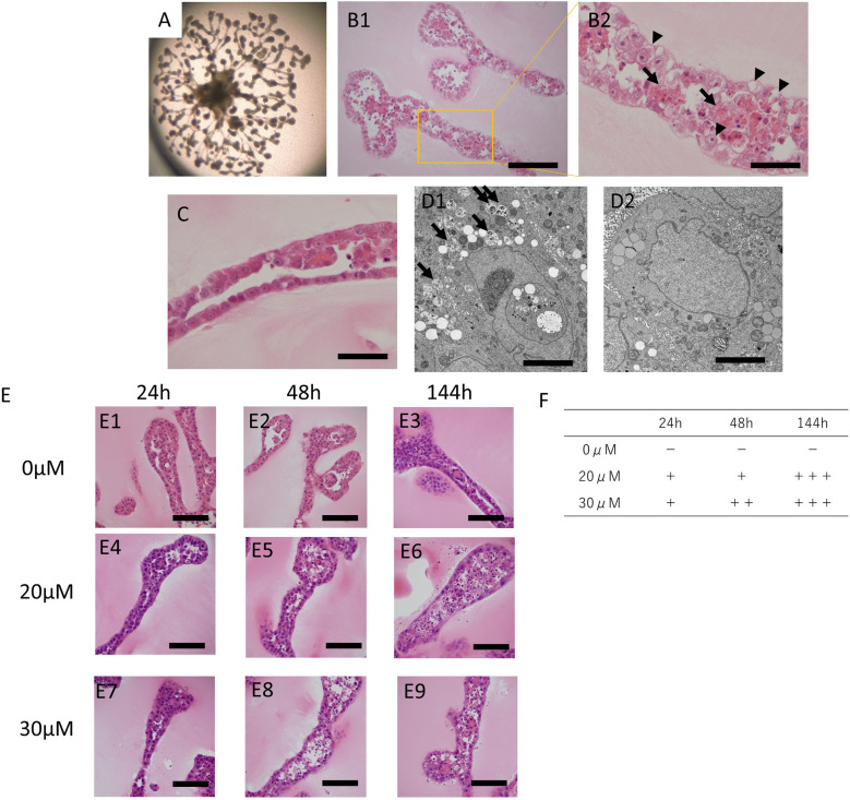
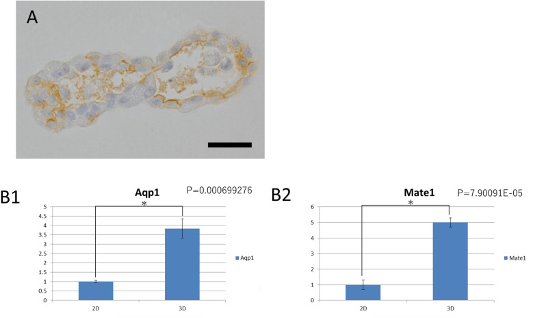
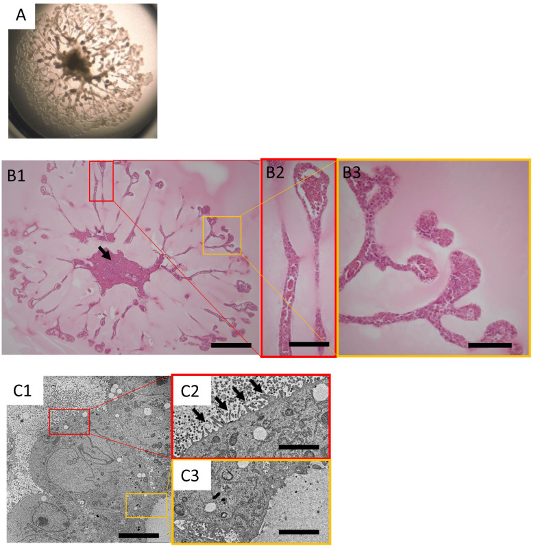
Organoids derived from renal tissue stem cells (KS cells) isolated from the S3 segment of adult rat nephrons have previously been developed and evaluated. However, data regarding the histopathological evaluation of these organoids are limited. Therefore, in this study, we performed histopathological examinations of the properties of these organoids and evaluated the nephrotoxicity changes induced by cisplatin treatment. We observe that the tubular structure of the organoids was generally lined by a single layer of cells, in concordance with previous findings. Microvilli were exclusively observed under electron microscopy on the luminal side of this tubular structure. Moreover, the luminal side of the tubular structure was positive for aquaporin-1 (Aqp1), a marker of the proximal renal tubule. Cisplatin treatment induced cell death and degeneration, including cytoplasmic vacuolation, in cells within the tubular structure of the organoids. Cisplatin toxicity is associated with the induction of γ-H2AX (a marker of DNA damage) and the drop of phospho-histone H3 (a marker of cell division) levels. During the nephrotoxicity assessment, the kidney organoids displayed various features similar to those of the natural kidney, suggesting that it is possible to use these organoids in predicting nephrotoxicity. The histological evaluation of the organoids in this study provides insights into the mechanisms underlying nephrotoxicity.
期刊介绍:
JTP is a scientific journal that publishes original studies in the field of toxicological pathology and in a wide variety of other related fields. The main scope of the journal is listed below.
Administrative Opinions of Policymakers and Regulatory Agencies
Adverse Events
Carcinogenesis
Data of A Predominantly Negative Nature
Drug-Induced Hematologic Toxicity
Embryological Pathology
High Throughput Pathology
Historical Data of Experimental Animals
Immunohistochemical Analysis
Molecular Pathology
Nomenclature of Lesions
Non-mammal Toxicity Study
Result or Lesion Induced by Chemicals of Which Names Hidden on Account of the Authors
Technology and Methodology Related to Toxicological Pathology
Tumor Pathology; Neoplasia and Hyperplasia
Ultrastructural Analysis
Use of Animal Models.


 分享
分享
 求助内容:
求助内容: 应助结果提醒方式:
 扫码关注我们
