{"title":"Simultaneous bilateral inflammatory choroidal neovascularization in a case of healed serpiginous-like choroiditis.","authors":"Gitanjli Sood, Ramanuj Samanta, Devesh Kumawat, Prateek Nishant","doi":"10.3205/oc000199","DOIUrl":null,"url":null,"abstract":"<p><strong>Objective: </strong>Inflammatory choroidal neovascularization (i-CNV) is an infrequent but sight-threatening complication of posterior uveitis. Although it can occur in a wide range of infectious and non-infectious uveitides, presence of simultaneous bilateral i-CNV is rare. In this report, we present a unique case of bilateral simultaneous i-CNV in a young patient of healed tubercular serpiginous-like choroiditis.</p><p><strong>Method: </strong>A 20-year-old male presented with recent worsening of vision in the right eye for one month. Fundus examination revealed bilateral multifocal healed choroiditis lesions with right eye tiny subfoveal hemorrhage raising the suspicion of an underlying choroidal neovascularization. Fundus fluorescein angiography and optical coherence tomography confirmed presence of choroidal neovascular membrane in both eyes.</p><p><strong>Result: </strong>Resolution of activity was noted in both eyes after bilateral sequential intravitreal bevacizumab injections.</p><p><strong>Conclusion: </strong>Inflammatory choroidal neovascularization may be seen in patients with healed tubercular serpiginous-like choroiditis, after a long period of quiescence. Simultaneous bilateral presentation is rare but possible, requiring mandatory multimodal imaging of both eyes under high index of suspicion. Early institution of anti-vascular endothelial growth factor may salvage optimum vision in such a scenario.</p>","PeriodicalId":73178,"journal":{"name":"GMS ophthalmology cases","volume":" ","pages":"Doc12"},"PeriodicalIF":0.0000,"publicationDate":"2022-05-20","publicationTypes":"Journal Article","fieldsOfStudy":null,"isOpenAccess":false,"openAccessPdf":"https://www.ncbi.nlm.nih.gov/pmc/articles/PMC9284432/pdf/","citationCount":"0","resultStr":null,"platform":"Semanticscholar","paperid":null,"PeriodicalName":"GMS ophthalmology cases","FirstCategoryId":"1085","ListUrlMain":"https://doi.org/10.3205/oc000199","RegionNum":0,"RegionCategory":null,"ArticlePicture":[],"TitleCN":null,"AbstractTextCN":null,"PMCID":null,"EPubDate":"2022/1/1 0:00:00","PubModel":"eCollection","JCR":"","JCRName":"","Score":null,"Total":0}
引用次数: 0
Abstract
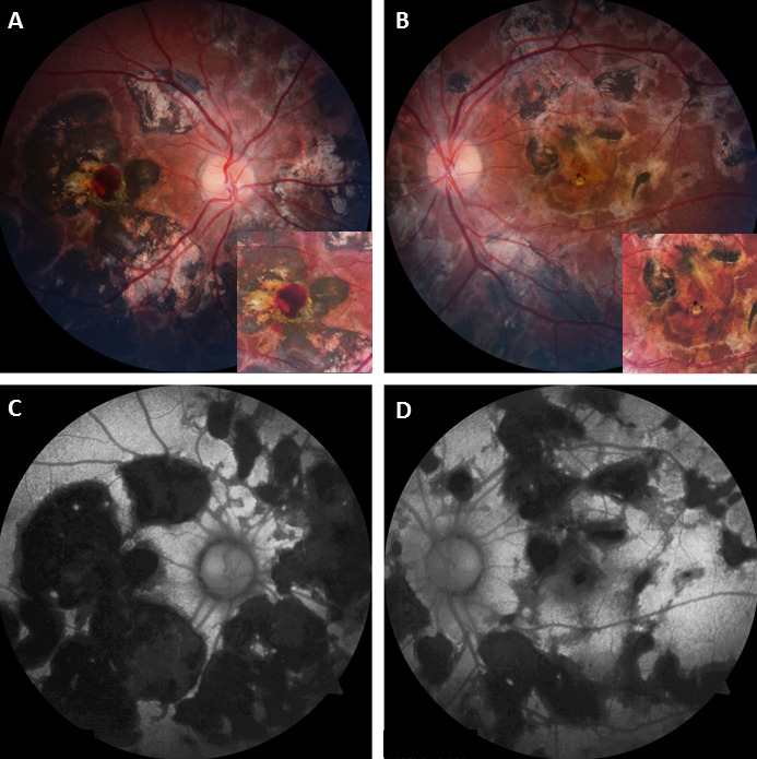
Objective: Inflammatory choroidal neovascularization (i-CNV) is an infrequent but sight-threatening complication of posterior uveitis. Although it can occur in a wide range of infectious and non-infectious uveitides, presence of simultaneous bilateral i-CNV is rare. In this report, we present a unique case of bilateral simultaneous i-CNV in a young patient of healed tubercular serpiginous-like choroiditis.
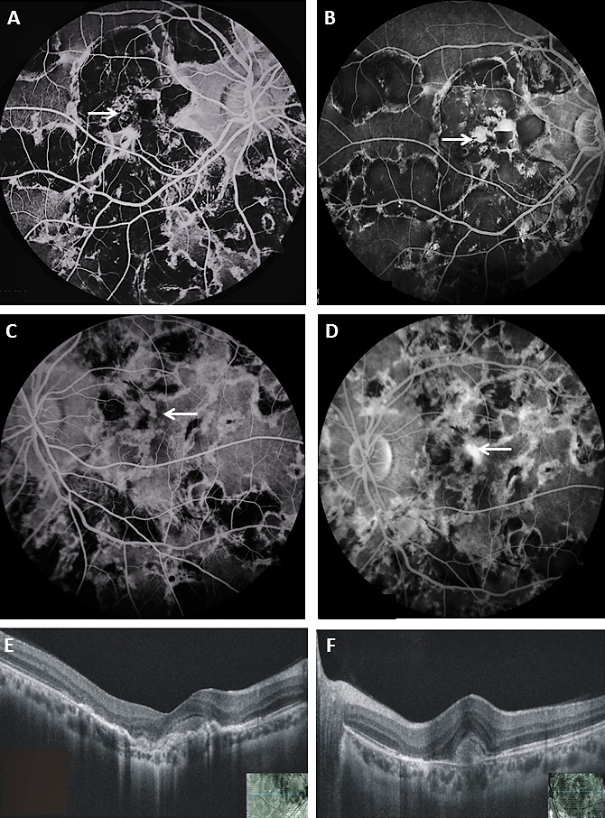
Method: A 20-year-old male presented with recent worsening of vision in the right eye for one month. Fundus examination revealed bilateral multifocal healed choroiditis lesions with right eye tiny subfoveal hemorrhage raising the suspicion of an underlying choroidal neovascularization. Fundus fluorescein angiography and optical coherence tomography confirmed presence of choroidal neovascular membrane in both eyes.
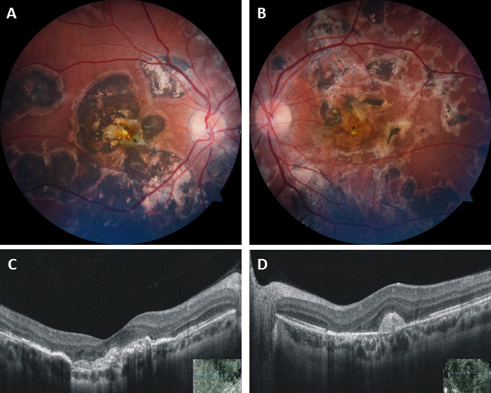
Result: Resolution of activity was noted in both eyes after bilateral sequential intravitreal bevacizumab injections.
Conclusion: Inflammatory choroidal neovascularization may be seen in patients with healed tubercular serpiginous-like choroiditis, after a long period of quiescence. Simultaneous bilateral presentation is rare but possible, requiring mandatory multimodal imaging of both eyes under high index of suspicion. Early institution of anti-vascular endothelial growth factor may salvage optimum vision in such a scenario.


 分享
分享

 求助内容:
求助内容: 应助结果提醒方式:
 扫码关注我们
