Bilge Batu Oto, Aslihan Yilmaz Çebi, Oguzhan Kiliçarslan, Ahmet Murat Sarici
{"title":"Multimodal imaging of a sporadic retinal astrocytic hamartoma simulating retinoblastoma in a newborn.","authors":"Bilge Batu Oto, Aslihan Yilmaz Çebi, Oguzhan Kiliçarslan, Ahmet Murat Sarici","doi":"10.3205/oc000198","DOIUrl":null,"url":null,"abstract":"<p><strong>Introduction: </strong>To report a sporadic astrocytic hamartoma simulating retinoblastoma in a newborn.</p><p><strong>Methods: </strong>Clinical data was reviewed retrospectively.</p><p><strong>Results: </strong>A 3-month-old baby with a history of perinatal asphyxia was referred to our ocular oncology clinic with suspected retinoblastoma in the left eye. Dilated fundoscopy revealed a solitary tumor covering the optic disc at the left eye. The whitish-yellow lesion was well-defined, opaque, and minimally calcified. High internal reflectivity and posterior shadowing due to the intralesional calcification, and intratumoral cystic spaces were observed in B-scan ultrasound imaging. Optical coherence tomography imaging showed an intraretinal tumor with cystic spaces and posterior shadowing. The tumor was diagnosed as an astrocytic hamartoma. The systemic evaluation was negative for phacomatoses. The lesion has been observed with multimodal imaging for six years without significant changes.</p><p><strong>Conclusions: </strong>Retinal astrocytic hamartomas are benign tumors that arise within the retinal nerve fiber layer. Differential diagnosis constitutes high importance since they may be misdiagnosed as retinoblastoma, and therefore may be overtreated. Whereas retinoblastoma requires immediate treatment, retinal astrocytic hamartomas are commonly followed-up. Multimodal imaging with B-scan ultrasonography and optical coherence tomography are useful in distinguishing those two entities.</p>","PeriodicalId":73178,"journal":{"name":"GMS ophthalmology cases","volume":" ","pages":"Doc11"},"PeriodicalIF":0.0000,"publicationDate":"2022-05-20","publicationTypes":"Journal Article","fieldsOfStudy":null,"isOpenAccess":false,"openAccessPdf":"https://www.ncbi.nlm.nih.gov/pmc/articles/PMC9284428/pdf/","citationCount":"0","resultStr":null,"platform":"Semanticscholar","paperid":null,"PeriodicalName":"GMS ophthalmology cases","FirstCategoryId":"1085","ListUrlMain":"https://doi.org/10.3205/oc000198","RegionNum":0,"RegionCategory":null,"ArticlePicture":[],"TitleCN":null,"AbstractTextCN":null,"PMCID":null,"EPubDate":"2022/1/1 0:00:00","PubModel":"eCollection","JCR":"","JCRName":"","Score":null,"Total":0}
引用次数: 0
Abstract
Introduction: To report a sporadic astrocytic hamartoma simulating retinoblastoma in a newborn.
Methods: Clinical data was reviewed retrospectively.
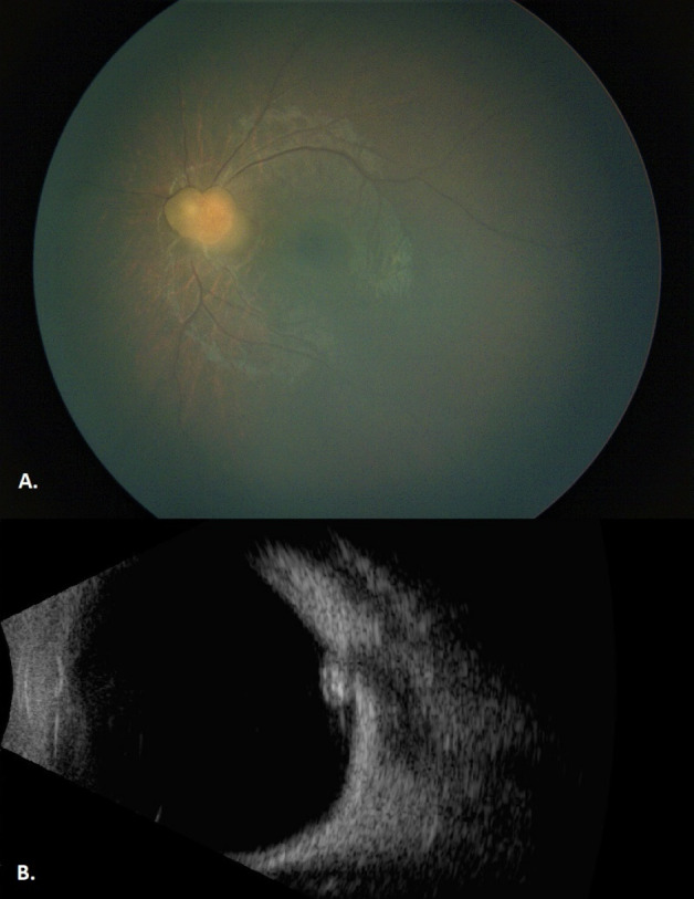
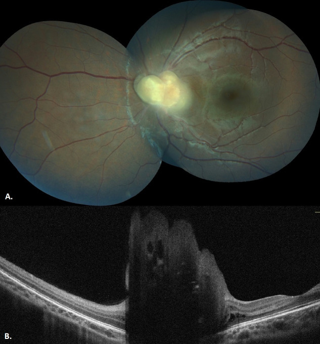
Results: A 3-month-old baby with a history of perinatal asphyxia was referred to our ocular oncology clinic with suspected retinoblastoma in the left eye. Dilated fundoscopy revealed a solitary tumor covering the optic disc at the left eye. The whitish-yellow lesion was well-defined, opaque, and minimally calcified. High internal reflectivity and posterior shadowing due to the intralesional calcification, and intratumoral cystic spaces were observed in B-scan ultrasound imaging. Optical coherence tomography imaging showed an intraretinal tumor with cystic spaces and posterior shadowing. The tumor was diagnosed as an astrocytic hamartoma. The systemic evaluation was negative for phacomatoses. The lesion has been observed with multimodal imaging for six years without significant changes.
Conclusions: Retinal astrocytic hamartomas are benign tumors that arise within the retinal nerve fiber layer. Differential diagnosis constitutes high importance since they may be misdiagnosed as retinoblastoma, and therefore may be overtreated. Whereas retinoblastoma requires immediate treatment, retinal astrocytic hamartomas are commonly followed-up. Multimodal imaging with B-scan ultrasonography and optical coherence tomography are useful in distinguishing those two entities.


 分享
分享

 求助内容:
求助内容: 应助结果提醒方式:
 扫码关注我们
