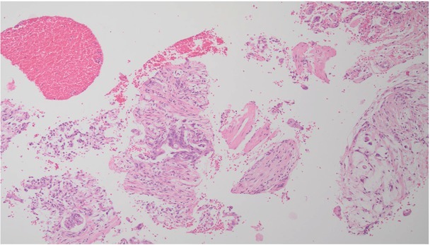
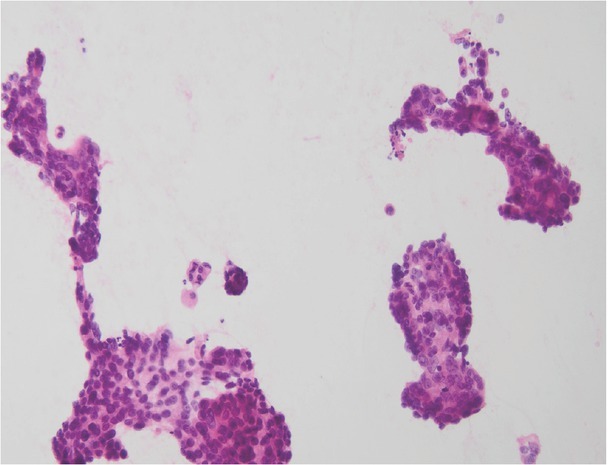
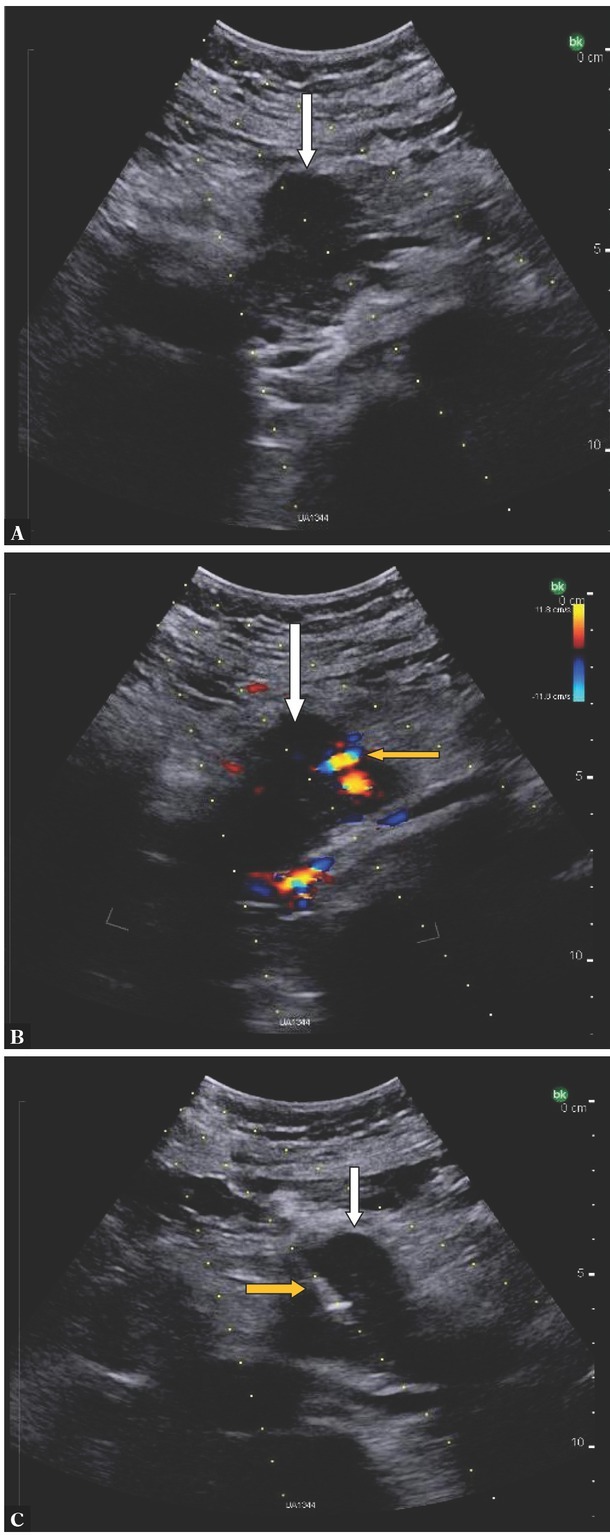
{"title":"Ultrasound-guided Percutaneous Core-needle Biopsy of Focal Pancreatic Lesions - Practical Aspectss.","authors":"Agnieszka Zofia Rogowska","doi":"10.15557/JoU.2022.0019","DOIUrl":null,"url":null,"abstract":"<p><p>Ultrasound-guided percutaneous core-needle biopsy is an excellent diagnostic tool for solid pancreatic lesions. It allows for identifying neoplastic pancreatic tumors with nearly 100% sensitivity, specificity and accuracy. Unresectable tumor assessment prior to planned palliative treatment is the primary indication for percutaneous pancreatic tumor biopsy. In the case of potential tumors eligible for radical surgery, endosonography-guided biopsy is used, if clinically necessary, to avoid the peritoneal spread of tumor cells during puncture. The possibility of obtaining a specimen for a detailed microscopic assessment during an easily accessible and simple procedure is the main advantage of core-needle biopsy over the low, yet higher when compared to other biopsy techniques, risk of complications. Obtaining tissue samples for molecular analysis is essential for palliative targeted therapy in pancreatic cancer and may become the main indication for the common core-needle biopsy of inoperable pancreatic tumors in the near future. The present paper describes the indications and the technique for core-needle biopsy in pancreatic tumors. Based on the studies published to date, the safety of the procedure, significant complications, including bleeding in particular, and the diagnostic sensitivity and specificity, also compared to other biopsy techniques, have been summarized. The present paper may contribute to the introduction of core-needle biopsy of pancreatic masses into clinical practice.</p>","PeriodicalId":45612,"journal":{"name":"Journal of Ultrasonography","volume":"22 89","pages":"117-120"},"PeriodicalIF":1.5000,"publicationDate":"2022-04-27","publicationTypes":"Journal Article","fieldsOfStudy":null,"isOpenAccess":false,"openAccessPdf":"https://ftp.ncbi.nlm.nih.gov/pub/pmc/oa_pdf/81/ab/jou-22-117.PMC9231516.pdf","citationCount":"2","resultStr":null,"platform":"Semanticscholar","paperid":null,"PeriodicalName":"Journal of Ultrasonography","FirstCategoryId":"1085","ListUrlMain":"https://doi.org/10.15557/JoU.2022.0019","RegionNum":0,"RegionCategory":null,"ArticlePicture":[],"TitleCN":null,"AbstractTextCN":null,"PMCID":null,"EPubDate":"2022/4/1 0:00:00","PubModel":"eCollection","JCR":"Q3","JCRName":"RADIOLOGY, NUCLEAR MEDICINE & MEDICAL IMAGING","Score":null,"Total":0}
引用次数: 2
Abstract
Ultrasound-guided percutaneous core-needle biopsy is an excellent diagnostic tool for solid pancreatic lesions. It allows for identifying neoplastic pancreatic tumors with nearly 100% sensitivity, specificity and accuracy. Unresectable tumor assessment prior to planned palliative treatment is the primary indication for percutaneous pancreatic tumor biopsy. In the case of potential tumors eligible for radical surgery, endosonography-guided biopsy is used, if clinically necessary, to avoid the peritoneal spread of tumor cells during puncture. The possibility of obtaining a specimen for a detailed microscopic assessment during an easily accessible and simple procedure is the main advantage of core-needle biopsy over the low, yet higher when compared to other biopsy techniques, risk of complications. Obtaining tissue samples for molecular analysis is essential for palliative targeted therapy in pancreatic cancer and may become the main indication for the common core-needle biopsy of inoperable pancreatic tumors in the near future. The present paper describes the indications and the technique for core-needle biopsy in pancreatic tumors. Based on the studies published to date, the safety of the procedure, significant complications, including bleeding in particular, and the diagnostic sensitivity and specificity, also compared to other biopsy techniques, have been summarized. The present paper may contribute to the introduction of core-needle biopsy of pancreatic masses into clinical practice.


 分享
分享
 求助内容:
求助内容: 应助结果提醒方式:
 扫码关注我们
