{"title":"Anaplastic Intraventricular Meningioma with Rhabdoid Features: An Unusual Tumor with Usual Clinical Presentation.","authors":"Preeti Agarwal, Nancy Gupta, Alok Srivastava, Madhu Kumar, Suarabh Kumar, Chhitij Srivastava","doi":"10.1177/2632010X221115157","DOIUrl":null,"url":null,"abstract":"<p><p>Meningiomas are tumors arising from leptomeninges. Malignant counterpart of them is known as anaplastic meningioma which are WHO grade III tumors. Intraventricular location of these tumors is rare and is clinic-radiologically challenging. Histopathology and immunohistochemistry are confirmatory. We present case of a 27-year-old girl, who presented with usual symptoms of intraventricular mass in emergency. After shunt surgery, clinical diagnosis of ependymoma was formed with differential of high-grade glioma. Squash tissue was difficult to crush displaying tight clusters of spindle cells with necrosis in background. Definitive histology revealed high grade spindle cell neoplasm disposed in sheets with brisk and atypical mitosis. Only focal whorling pattern was seen. Large cells with eccentric cytoplasm, reminiscent of rhabdoid cells were also seen. Immunohistochemistry was positive for vimentin and EMA, negative for GFAP. Final diagnosis of Anaplastic meningioma was dispatched. The histological pattern of the present case, young age of presentation and presence of Rhabdoid cells make it unusual. Though rare but intraventricular meningiomas must also be kept in clinical radiological differentials apart from the usual ependymoma at this location.</p>","PeriodicalId":53204,"journal":{"name":"Clinical Pathology","volume":" ","pages":"2632010X221115157"},"PeriodicalIF":1.9000,"publicationDate":"2022-07-29","publicationTypes":"Journal Article","fieldsOfStudy":null,"isOpenAccess":false,"openAccessPdf":"https://ftp.ncbi.nlm.nih.gov/pub/pmc/oa_pdf/48/bb/10.1177_2632010X221115157.PMC9340328.pdf","citationCount":"0","resultStr":null,"platform":"Semanticscholar","paperid":null,"PeriodicalName":"Clinical Pathology","FirstCategoryId":"1085","ListUrlMain":"https://doi.org/10.1177/2632010X221115157","RegionNum":0,"RegionCategory":null,"ArticlePicture":[],"TitleCN":null,"AbstractTextCN":null,"PMCID":null,"EPubDate":"2022/1/1 0:00:00","PubModel":"eCollection","JCR":"Q3","JCRName":"PATHOLOGY","Score":null,"Total":0}
引用次数: 0
Abstract
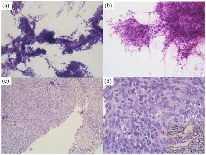
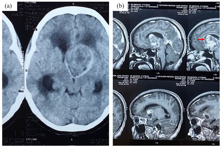
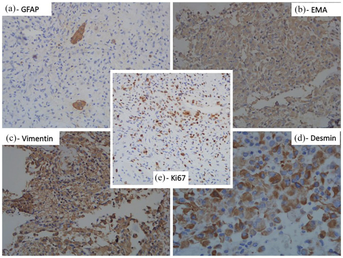
Meningiomas are tumors arising from leptomeninges. Malignant counterpart of them is known as anaplastic meningioma which are WHO grade III tumors. Intraventricular location of these tumors is rare and is clinic-radiologically challenging. Histopathology and immunohistochemistry are confirmatory. We present case of a 27-year-old girl, who presented with usual symptoms of intraventricular mass in emergency. After shunt surgery, clinical diagnosis of ependymoma was formed with differential of high-grade glioma. Squash tissue was difficult to crush displaying tight clusters of spindle cells with necrosis in background. Definitive histology revealed high grade spindle cell neoplasm disposed in sheets with brisk and atypical mitosis. Only focal whorling pattern was seen. Large cells with eccentric cytoplasm, reminiscent of rhabdoid cells were also seen. Immunohistochemistry was positive for vimentin and EMA, negative for GFAP. Final diagnosis of Anaplastic meningioma was dispatched. The histological pattern of the present case, young age of presentation and presence of Rhabdoid cells make it unusual. Though rare but intraventricular meningiomas must also be kept in clinical radiological differentials apart from the usual ependymoma at this location.


 分享
分享
 求助内容:
求助内容: 应助结果提醒方式:
 扫码关注我们
