Alexandra Lucaciu, Patrick Felix Samp, Elke Hattingen, Roxane-Isabelle Kestner, Petra Davidova, Thomas Kohnen, Jasmin Rudolph, Andreas Dietz, Helmuth Steinmetz, Adam Strzelczyk
{"title":"Sudden vision loss and neurological deficits after facial hyaluronic acid filler injection.","authors":"Alexandra Lucaciu, Patrick Felix Samp, Elke Hattingen, Roxane-Isabelle Kestner, Petra Davidova, Thomas Kohnen, Jasmin Rudolph, Andreas Dietz, Helmuth Steinmetz, Adam Strzelczyk","doi":"10.1186/s42466-022-00203-x","DOIUrl":null,"url":null,"abstract":"<p><strong>Background: </strong>The ongoing expansion of the cosmetic armamentarium of facial rejuvenation fails to uncover the inherent risks of cosmetic interventions. Informed consent to all risks of cosmetic filler injections and potential sequelae, including ocular and neurological complications, should be carefully ensured. We present two cases of complications following facial hyaluronic acid filler injections.</p><p><strong>Case presentations: </strong>Case 1: A 43-year-old woman presented with monocular vision loss of the left eye, associated ptosis, ophthalmoplegia, periocular pain and nausea, cutaneous changes of the glabella region and forehead, and sensory impairment in the left maxillary branch dermatome (V2) after receiving a hyaluronic acid (HA) filler injection into the left glabellar area. On ophthalmological examination, an ophthalmic artery occlusion (OAO) was diagnosed upon identification of a \"cherry-red spot\". Magnetic resonance imaging (MRI) revealed a left ischemic optic neuropathy. Supportive therapy and hyaluronidase injections were initiated. A follow-up MRI of the head performed two months after presentation corresponded to stable MRI findings. The patient had irreversible and complete vision loss of the left eye, however, the ptosis resolved. Case 2: A 29-year-old woman was admitted to hospital a few hours after a rhinoplasty and cheek augmentation with hyaluronic acid, presenting with acute monocular vision loss in the right eye, retrobulbar pain, fatigue and vomiting. In addition, the patient presented a harbinger of impending skin necrosis and a complete oculomotor nerve palsy on the right side, choroidal ischemia and vision impairment. Supportive treatment and hyaluronidase injections into the ischemic tissue were initiated. A small scar at the tip of the nose, vision impairment and an irregular pupillary margin on the right side persisted at follow-up.</p><p><strong>Conclusion: </strong>These two case reports and the literature review emphasize the pathophysiological mechanisms leading to potentially devastating complications. In order to reduce the risk of vision loss secondary to cosmetic filler injections, practitioners should possess a thorough knowledge of anatomy and preventive strategies.</p>","PeriodicalId":19169,"journal":{"name":"Neurological Research and Practice","volume":" ","pages":"40"},"PeriodicalIF":0.0000,"publicationDate":"2022-07-18","publicationTypes":"Journal Article","fieldsOfStudy":null,"isOpenAccess":false,"openAccessPdf":"https://www.ncbi.nlm.nih.gov/pmc/articles/PMC9290300/pdf/","citationCount":"6","resultStr":null,"platform":"Semanticscholar","paperid":null,"PeriodicalName":"Neurological Research and Practice","FirstCategoryId":"1085","ListUrlMain":"https://doi.org/10.1186/s42466-022-00203-x","RegionNum":0,"RegionCategory":null,"ArticlePicture":[],"TitleCN":null,"AbstractTextCN":null,"PMCID":null,"EPubDate":"","PubModel":"","JCR":"","JCRName":"","Score":null,"Total":0}
引用次数: 6
Abstract
Background: The ongoing expansion of the cosmetic armamentarium of facial rejuvenation fails to uncover the inherent risks of cosmetic interventions. Informed consent to all risks of cosmetic filler injections and potential sequelae, including ocular and neurological complications, should be carefully ensured. We present two cases of complications following facial hyaluronic acid filler injections.
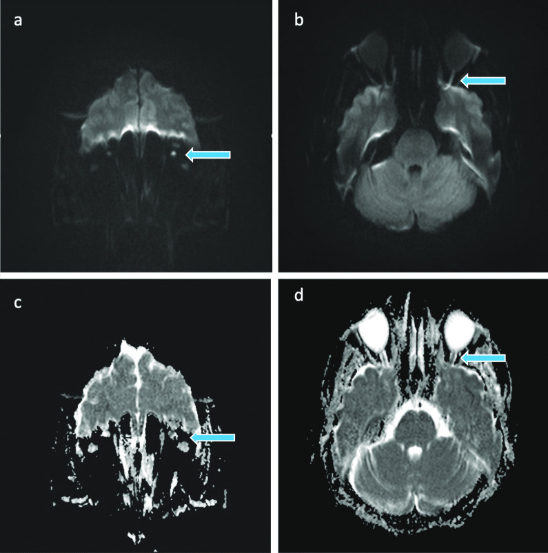
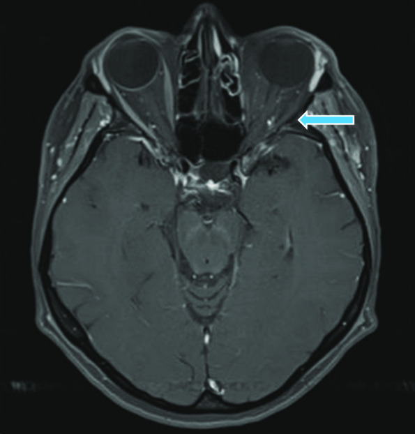
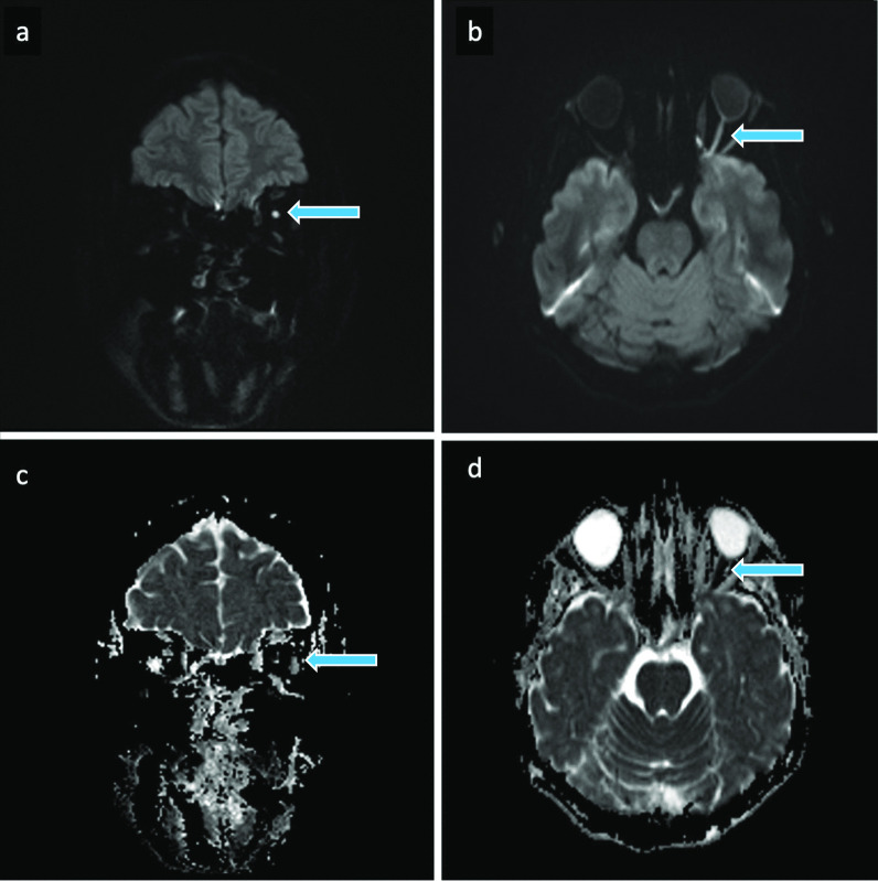
Case presentations: Case 1: A 43-year-old woman presented with monocular vision loss of the left eye, associated ptosis, ophthalmoplegia, periocular pain and nausea, cutaneous changes of the glabella region and forehead, and sensory impairment in the left maxillary branch dermatome (V2) after receiving a hyaluronic acid (HA) filler injection into the left glabellar area. On ophthalmological examination, an ophthalmic artery occlusion (OAO) was diagnosed upon identification of a "cherry-red spot". Magnetic resonance imaging (MRI) revealed a left ischemic optic neuropathy. Supportive therapy and hyaluronidase injections were initiated. A follow-up MRI of the head performed two months after presentation corresponded to stable MRI findings. The patient had irreversible and complete vision loss of the left eye, however, the ptosis resolved. Case 2: A 29-year-old woman was admitted to hospital a few hours after a rhinoplasty and cheek augmentation with hyaluronic acid, presenting with acute monocular vision loss in the right eye, retrobulbar pain, fatigue and vomiting. In addition, the patient presented a harbinger of impending skin necrosis and a complete oculomotor nerve palsy on the right side, choroidal ischemia and vision impairment. Supportive treatment and hyaluronidase injections into the ischemic tissue were initiated. A small scar at the tip of the nose, vision impairment and an irregular pupillary margin on the right side persisted at follow-up.
Conclusion: These two case reports and the literature review emphasize the pathophysiological mechanisms leading to potentially devastating complications. In order to reduce the risk of vision loss secondary to cosmetic filler injections, practitioners should possess a thorough knowledge of anatomy and preventive strategies.


 分享
分享

 求助内容:
求助内容: 应助结果提醒方式:
 扫码关注我们
