Ritica Chaudhary, Mona Lisa, Payal Kumari, Aman Kumar
{"title":"Dedifferentiated Liposarcoma: A Rare Case Report of Retroperitoneal Myxoid Soft Tissue Tumour with Diagnostic Dilemma.","authors":"Ritica Chaudhary, Mona Lisa, Payal Kumari, Aman Kumar","doi":"10.1177/2632010X221112455","DOIUrl":null,"url":null,"abstract":"<p><strong>Background: </strong>The retroperitoneum can host a wide spectrum of soft tissue lesions. These tumours pose a challenge to the pathologist as the morphology is not of much help and immunohistochemistry becomes a necessity.</p><p><strong>Case report: </strong>Sixty years old male presented with 2 months history of abdominal lump, pain and dyspepsia. The MRI revealed a heterogeneous mass in the retroperitoneum involving right para spinal muscle, right iliac fossa and right perinephric region with destruction of right transverse process and erosion of adjacent L3 vertebra. Trucut biopsy of the mass was reported as fibroliposarcoma at an outside lab. Patient underwent a wide local excision. Grossly the tumour gave an impression of a liposarcoma but the microscopy showed areas of spindle cells, epitheloid cells, focal areas of ganglion like cells and large areas of myxoid change. IHC panel of S-100, SMA, caldesmon, myogenin, myoglobin and Alk-1 was negative. MDM2, CDK4 and p16 IHC came positive proving it to be a dedifferentiated liposarcoma.</p><p><strong>Conclusion: </strong>We report a curious case of retroperitoneal soft tissue tumour with complex morphology and IHC features diagnosed as dedifferentiated liposarcoma based on MDM2, CDK and p16 positivity.</p>","PeriodicalId":53204,"journal":{"name":"Clinical Pathology","volume":" ","pages":"2632010X221112455"},"PeriodicalIF":1.9000,"publicationDate":"2022-07-19","publicationTypes":"Journal Article","fieldsOfStudy":null,"isOpenAccess":false,"openAccessPdf":"https://www.ncbi.nlm.nih.gov/pmc/articles/PMC9301116/pdf/","citationCount":"0","resultStr":null,"platform":"Semanticscholar","paperid":null,"PeriodicalName":"Clinical Pathology","FirstCategoryId":"1085","ListUrlMain":"https://doi.org/10.1177/2632010X221112455","RegionNum":0,"RegionCategory":null,"ArticlePicture":[],"TitleCN":null,"AbstractTextCN":null,"PMCID":null,"EPubDate":"2022/1/1 0:00:00","PubModel":"eCollection","JCR":"Q3","JCRName":"PATHOLOGY","Score":null,"Total":0}
引用次数: 0
Abstract
Background: The retroperitoneum can host a wide spectrum of soft tissue lesions. These tumours pose a challenge to the pathologist as the morphology is not of much help and immunohistochemistry becomes a necessity.
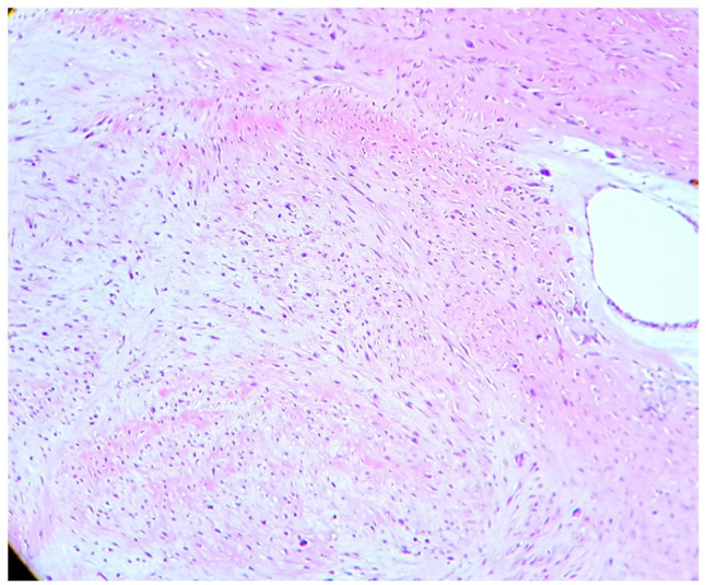
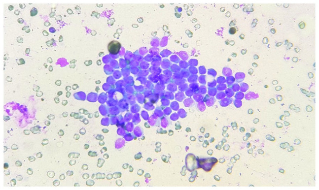
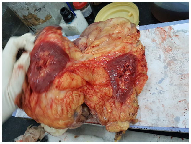
Case report: Sixty years old male presented with 2 months history of abdominal lump, pain and dyspepsia. The MRI revealed a heterogeneous mass in the retroperitoneum involving right para spinal muscle, right iliac fossa and right perinephric region with destruction of right transverse process and erosion of adjacent L3 vertebra. Trucut biopsy of the mass was reported as fibroliposarcoma at an outside lab. Patient underwent a wide local excision. Grossly the tumour gave an impression of a liposarcoma but the microscopy showed areas of spindle cells, epitheloid cells, focal areas of ganglion like cells and large areas of myxoid change. IHC panel of S-100, SMA, caldesmon, myogenin, myoglobin and Alk-1 was negative. MDM2, CDK4 and p16 IHC came positive proving it to be a dedifferentiated liposarcoma.
Conclusion: We report a curious case of retroperitoneal soft tissue tumour with complex morphology and IHC features diagnosed as dedifferentiated liposarcoma based on MDM2, CDK and p16 positivity.


 分享
分享
 求助内容:
求助内容: 应助结果提醒方式:
 扫码关注我们
