{"title":"Retroperitoneal Liposarcoma: The Giant Type.","authors":"Subhi Mansour, Nassim Azzam, Yoram Kluger, Safi Khuri","doi":"10.14740/jmc4014","DOIUrl":null,"url":null,"abstract":"<p><p>Retroperitoneal tumors (RPTs) are very rare tumors that arise in the large space of the retroperitoneum. About two-third of these tumors are malignant, of which soft tissue sarcoma (STS) is the most common and comprises almost one-third of malignant RPTs. Twenty to thirty percent of RPTs are benign. The retroperitoneal cavity has a very large potential space for tumor enlargement to a very high diameters without causing specific symptoms, especially during the initial phase of tumor enlargement. On diagnosis, the average tumor weight is 15 - 20 kg and tumor diameter is 20 - 25 cm. The most common retroperitoneal sarcoma type is liposarcoma, which account for 20% of all sarcoma types and 40% of all retroperitoneal sarcomas (RPSs). Other less common STS arise in the retroperitoneum include leiomyosarcoma and undifferentiated pleomorphic type. Giant liposarcoma is usually defined either as tumor diameter of 30 cm or more or tumor weight of 20 kg or higher. This specific type of sarcoma is very uncommon, with few case reports published in the English literature. Herein, we present a case of a healthy 33-year-old male patient, who was admitted due to abdominal distension and increased body weight since few months. An abdominopelvic computed tomography (CT) scan demonstrated a giant retroperitoneal mass of almost 40 cm in diameter in its largest dimension, located in the right retroperitoneal space. Ultrasound (US)-guided fine needle biopsy (FNB) was consistent with well differentiated liposarcoma. Surgical resection of the tumor along with the right colon, right ureter and kidney, third and fourth duodenal parts and part of the right iliopsoas muscle was contemplated. Histopathological report revealed well-differentiated liposarcoma of 50 cm in diameter, with foci of dedifferentiation, presented by pleomorphic sarcoma. Surgical margins were microscopically negative.</p>","PeriodicalId":16279,"journal":{"name":"Journal of Medical Cases","volume":"13 10","pages":"517-520"},"PeriodicalIF":0.0000,"publicationDate":"2022-10-01","publicationTypes":"Journal Article","fieldsOfStudy":null,"isOpenAccess":false,"openAccessPdf":"https://ftp.ncbi.nlm.nih.gov/pub/pmc/oa_pdf/1a/0d/jmc-13-517.PMC9635766.pdf","citationCount":"1","resultStr":null,"platform":"Semanticscholar","paperid":null,"PeriodicalName":"Journal of Medical Cases","FirstCategoryId":"1085","ListUrlMain":"https://doi.org/10.14740/jmc4014","RegionNum":0,"RegionCategory":null,"ArticlePicture":[],"TitleCN":null,"AbstractTextCN":null,"PMCID":null,"EPubDate":"2022/10/31 0:00:00","PubModel":"Epub","JCR":"","JCRName":"","Score":null,"Total":0}
引用次数: 1
Abstract
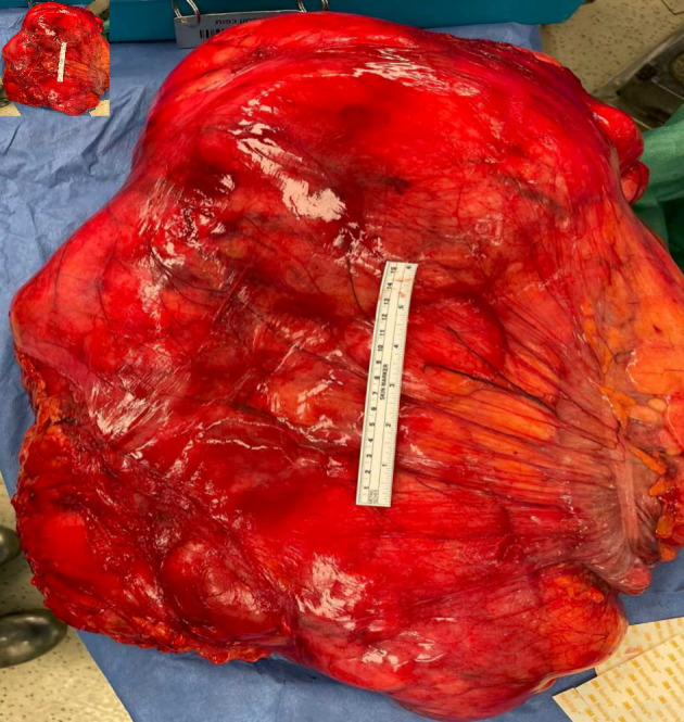
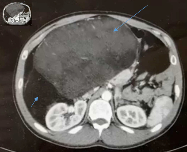
Retroperitoneal tumors (RPTs) are very rare tumors that arise in the large space of the retroperitoneum. About two-third of these tumors are malignant, of which soft tissue sarcoma (STS) is the most common and comprises almost one-third of malignant RPTs. Twenty to thirty percent of RPTs are benign. The retroperitoneal cavity has a very large potential space for tumor enlargement to a very high diameters without causing specific symptoms, especially during the initial phase of tumor enlargement. On diagnosis, the average tumor weight is 15 - 20 kg and tumor diameter is 20 - 25 cm. The most common retroperitoneal sarcoma type is liposarcoma, which account for 20% of all sarcoma types and 40% of all retroperitoneal sarcomas (RPSs). Other less common STS arise in the retroperitoneum include leiomyosarcoma and undifferentiated pleomorphic type. Giant liposarcoma is usually defined either as tumor diameter of 30 cm or more or tumor weight of 20 kg or higher. This specific type of sarcoma is very uncommon, with few case reports published in the English literature. Herein, we present a case of a healthy 33-year-old male patient, who was admitted due to abdominal distension and increased body weight since few months. An abdominopelvic computed tomography (CT) scan demonstrated a giant retroperitoneal mass of almost 40 cm in diameter in its largest dimension, located in the right retroperitoneal space. Ultrasound (US)-guided fine needle biopsy (FNB) was consistent with well differentiated liposarcoma. Surgical resection of the tumor along with the right colon, right ureter and kidney, third and fourth duodenal parts and part of the right iliopsoas muscle was contemplated. Histopathological report revealed well-differentiated liposarcoma of 50 cm in diameter, with foci of dedifferentiation, presented by pleomorphic sarcoma. Surgical margins were microscopically negative.


 分享
分享

 求助内容:
求助内容: 应助结果提醒方式:
 扫码关注我们
