Retinal neurodegeneration in eyes with NPDR risk phenotypes: A two-year longitudinal study

IF 3 3区 医学 Q1 OPHTHALMOLOGY Acta Ophthalmologica Pub Date : 2023-10-05 DOI:10.1111/aos.15787
Débora Reste-Ferreira, Inês Pereira Marques, Torcato Santos, Maria Luísa Ribeiro, Luís Mendes, Ana Rita Santos, Conceição Lobo, José Cunha-Vaz
{"title":"Retinal neurodegeneration in eyes with NPDR risk phenotypes: A two-year longitudinal study","authors":"Débora Reste-Ferreira,&nbsp;Inês Pereira Marques,&nbsp;Torcato Santos,&nbsp;Maria Luísa Ribeiro,&nbsp;Luís Mendes,&nbsp;Ana Rita Santos,&nbsp;Conceição Lobo,&nbsp;José Cunha-Vaz","doi":"10.1111/aos.15787","DOIUrl":null,"url":null,"abstract":"<div>\n \n \n <section>\n \n <h3> Introduction</h3>\n \n <p>Diabetic retinopathy (DR) is both a microangiopathy and a neurodegenerative disease. However, the connections between both changes are not well known.</p>\n </section>\n \n <section>\n \n <h3> Purpose</h3>\n \n <p>To characterise the longitudinal retinal ganglion cell layer + inner plexiform layer (GCL + IPL) changes and their association with microvascular changes in type-2 diabetes (T2D) patients with nonproliferative diabetic retinopathy (NPDR).</p>\n </section>\n \n <section>\n \n <h3> Methods</h3>\n \n <p>This two-year prospective study (CORDIS, NCT03696810) included 122 T2D individuals with NPDR identified as risk phenotypes B and C, which present a more rapid progression. Phenotype C was identified by decreased VD ≥ 2SD in healthy controls, and phenotype B, identified by subclinical macular oedema with only minimal vascular closure. The GCL + IPL thickness, vessel density, perfusion density and area of intercapillary spaces (AIS) were assessed by optical coherence tomography (OCT) and OCT angiography (OCTA). Linear mixed effects models were employed to evaluate the retinal GCL + IPL progression and its associations.</p>\n </section>\n \n <section>\n \n <h3> Results</h3>\n \n <p>Regarding GCL + IPL thickness, T2D individuals presented on average 80.1 ± 7.49 μm, statistically significantly lower than the healthy control group, 82.5 ± 5.71 (<i>p</i> = 0.022), with only phenotype C differing significantly from controls (<i>p</i> = 0.006). GCL + IPL thickness steadily decreased during the two-year period in both risk phenotypes, with an annual decline rate of −0.372 μm/year (<i>p</i> &lt; 0.001). Indeed, phenotype C showed a higher rate of progression (−0.459 μm/year, <i>p</i> &lt; 0.001) when compared to phenotype B (−0.296 μm/year, <i>p</i> = 0.036). Eyes with ETDRS grade 20 showed GCL + IPL thickness values comparable to those of healthy control group (83.3 ± 5.80 and 82.7 ± 5.50 μm, respectively, <i>p</i> = 0.880), whereas there was a progressive decrease in GCL + IPL thickness in ETDRS grades 35 and 43–47 associated with the increase in severity of the retinopathy (−0.276 μm/year, <i>p</i> = 0.004; −0.585 μm/year, <i>p</i> = 0.013, respectively). Furthermore, the study showed statistically significant associations between the progressive thinning of GCL + IPL and the progressive increase in retinal capillary non-perfusion, with particular relevance for AIS (<i>p</i> &lt; 0.001).</p>\n </section>\n \n <section>\n \n <h3> Conclusions</h3>\n \n <p>Our findings showed that, in eyes with NPDR and at risk for progression, retinal neurodegeneration occurs at different rates in different risk phenotypes, and it is associated with retinal microvascular non-perfusion.</p>\n </section>\n </div>","PeriodicalId":6915,"journal":{"name":"Acta Ophthalmologica","volume":"102 4","pages":"e539-e547"},"PeriodicalIF":3.0000,"publicationDate":"2023-10-05","publicationTypes":"Journal Article","fieldsOfStudy":null,"isOpenAccess":false,"openAccessPdf":"https://onlinelibrary.wiley.com/doi/epdf/10.1111/aos.15787","citationCount":"0","resultStr":null,"platform":"Semanticscholar","paperid":null,"PeriodicalName":"Acta Ophthalmologica","FirstCategoryId":"3","ListUrlMain":"https://onlinelibrary.wiley.com/doi/10.1111/aos.15787","RegionNum":3,"RegionCategory":"医学","ArticlePicture":[],"TitleCN":null,"AbstractTextCN":null,"PMCID":null,"EPubDate":"","PubModel":"","JCR":"Q1","JCRName":"OPHTHALMOLOGY","Score":null,"Total":0}
引用次数: 0

Abstract

Introduction

Diabetic retinopathy (DR) is both a microangiopathy and a neurodegenerative disease. However, the connections between both changes are not well known.

Purpose

To characterise the longitudinal retinal ganglion cell layer + inner plexiform layer (GCL + IPL) changes and their association with microvascular changes in type-2 diabetes (T2D) patients with nonproliferative diabetic retinopathy (NPDR).

Methods

This two-year prospective study (CORDIS, NCT03696810) included 122 T2D individuals with NPDR identified as risk phenotypes B and C, which present a more rapid progression. Phenotype C was identified by decreased VD ≥ 2SD in healthy controls, and phenotype B, identified by subclinical macular oedema with only minimal vascular closure. The GCL + IPL thickness, vessel density, perfusion density and area of intercapillary spaces (AIS) were assessed by optical coherence tomography (OCT) and OCT angiography (OCTA). Linear mixed effects models were employed to evaluate the retinal GCL + IPL progression and its associations.

Results

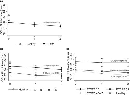
Regarding GCL + IPL thickness, T2D individuals presented on average 80.1 ± 7.49 μm, statistically significantly lower than the healthy control group, 82.5 ± 5.71 (p = 0.022), with only phenotype C differing significantly from controls (p = 0.006). GCL + IPL thickness steadily decreased during the two-year period in both risk phenotypes, with an annual decline rate of −0.372 μm/year (p < 0.001). Indeed, phenotype C showed a higher rate of progression (−0.459 μm/year, p < 0.001) when compared to phenotype B (−0.296 μm/year, p = 0.036). Eyes with ETDRS grade 20 showed GCL + IPL thickness values comparable to those of healthy control group (83.3 ± 5.80 and 82.7 ± 5.50 μm, respectively, p = 0.880), whereas there was a progressive decrease in GCL + IPL thickness in ETDRS grades 35 and 43–47 associated with the increase in severity of the retinopathy (−0.276 μm/year, p = 0.004; −0.585 μm/year, p = 0.013, respectively). Furthermore, the study showed statistically significant associations between the progressive thinning of GCL + IPL and the progressive increase in retinal capillary non-perfusion, with particular relevance for AIS (p < 0.001).

Conclusions

Our findings showed that, in eyes with NPDR and at risk for progression, retinal neurodegeneration occurs at different rates in different risk phenotypes, and it is associated with retinal microvascular non-perfusion.
Abstract Image

查看原文
分享 分享
微信好友 朋友圈 QQ好友 复制链接
本刊更多论文
具有NPDR风险表型的眼睛的视网膜神经退行性变:一项为期两年的纵向研究。
引言:糖尿病视网膜病变(DR)既是一种微血管病,也是一种神经退行性疾病。然而,这两种变化之间的联系尚不清楚。目的:表征视网膜纵向神经节细胞层 + 内部丛状层 + IPL)变化及其与2型糖尿病(T2D)非增殖性糖尿病视网膜病变(NPDR)患者微血管变化的关系。方法:这项为期两年的前瞻性研究(CORDIS,NCT03696810)纳入了122名具有NPDR风险表型B和C的T2D患者,其进展更快。表型C通过VD降低来鉴定 ≥ 健康对照中的2SD,以及表型B,通过亚临床黄斑水肿鉴定,只有最小的血管闭合。GCL + 通过光学相干断层扫描(OCT)和OCT血管造影术(OCTA)评估IPL厚度、血管密度、灌注密度和毛细血管间隙面积(AIS)。采用线性混合效应模型评估视网膜GCL + IPL进展及其相关性。结果:关于GCL + IPL厚度,T2D患者平均80.1 ± 7.49 μm,统计学上显著低于健康对照组82.5 ± 5.71(p = 0.022),只有表型C与对照组有显著差异(p = 0.006).GCL + 在两年期间,两种风险表型的IPL厚度均稳步下降,年下降率为-0.372 μm/年(p 结论:我们的研究结果表明,在NPDR患者和有进展风险的眼睛中,不同风险表型的视网膜神经退行性变发生率不同,并且与视网膜微血管非灌注有关。
本文章由计算机程序翻译,如有差异,请以英文原文为准。
求助全文
约1分钟内获得全文 去求助
来源期刊
Acta Ophthalmologica
Acta Ophthalmologica 医学-眼科学
CiteScore
7.60
自引率
5.90%
发文量
433
审稿时长
6 months
期刊介绍: Acta Ophthalmologica is published on behalf of the Acta Ophthalmologica Scandinavica Foundation and is the official scientific publication of the following societies: The Danish Ophthalmological Society, The Finnish Ophthalmological Society, The Icelandic Ophthalmological Society, The Norwegian Ophthalmological Society and The Swedish Ophthalmological Society, and also the European Association for Vision and Eye Research (EVER). Acta Ophthalmologica publishes clinical and experimental original articles, reviews, editorials, educational photo essays (Diagnosis and Therapy in Ophthalmology), case reports and case series, letters to the editor and doctoral theses.
期刊最新文献
Efficacy of intravitreal faricimab therapy for polypoidal choroidal vasculopathy: A systematic review and meta-analysis. The ever ongoing cosmetic quest to change eye colour. Slowing myopia progression with cylindrical annular refractive elements (CARE) spectacle lenses-Year 1 results from a 2-year prospective, multi-centre trial. Correlation of retinal fluid and photoreceptor and RPE loss in neovascular AMD by automated quantification, a real-world FRB! analysis. Incidence, risk factors, and patient characteristics in severe contact lens-related microbial keratitis.
×
引用
GB/T 7714-2015
复制
MLA
复制
APA
复制
导出至
BibTeX EndNote RefMan NoteFirst NoteExpress
×
×
提示
您的信息不完整,为了账户安全,请先补充。
现在去补充
×
提示
您因"违规操作"
具体请查看互助需知
我知道了
×
提示
现在去查看 取消
×
提示
确定
0
微信
客服QQ
Book学术公众号 扫码关注我们
反馈
×
意见反馈
请填写您的意见或建议
请填写您的手机或邮箱
已复制链接
已复制链接
快去分享给好友吧!
我知道了
×
扫码分享
扫码分享
Book学术官方微信
Book学术文献互助
Book学术文献互助群
群 号:481959085
Book学术
文献互助 智能选刊 最新文献 互助须知 联系我们:info@booksci.cn
Book学术提供免费学术资源搜索服务,方便国内外学者检索中英文文献。致力于提供最便捷和优质的服务体验。
Copyright © 2023 Book学术 All rights reserved.
ghs 京公网安备 11010802042870号 京ICP备2023020795号-1