{"title":"A case of radicular cyst on deciduous tooth in a 7-year-old child.","authors":"Takaaki Oda, Masanori Takada, Junya Ono, Yoriaki Kanri, Yasuo Okada, Ichiro Ogura","doi":"10.1007/s11282-023-00714-6","DOIUrl":null,"url":null,"abstract":"<p><p>Radicular cysts are the most common cystic lesions in the oral cavity, and have a rare occurrence in the primary dentition. We report a case of radicular cyst of mandible in child by multimodal imaging including panoramic radiography, CT, and MR imaging. A 7-year-old girl presented with swelling and without pain, and hypoesthesia on the right side of the mandible. On clinical examination, an expansive lesion with undulation was found to the buccal cortex of the right side of the mandible. Panoramic radiograph showed a unilocular radiolucency with well-defined margin, displaced tooth, and root resorption in the right mandible. Regarding CT imaging, axial soft tissue algorithm CT and bone tissue algorithm CT showed a low-attenuation internal structure and expansion of the buccal cortex of the right side of the mandible. Three-dimensional-CT showed expansion of the buccal cortex of the right side of the mandible. Multiplanar reformation imaging showed displaced tooth, root resorption, and expansion of the buccal cortex of the right side of the mandible. On T1-weighted image, the expansive lesion showed low signal intensity, and T2-weighted and STIR images revealed high signal intensity. A partial biopsy of the mandibular region was performed. Histopathological diagnosis was radicular cyst caused by apical periodontitis with abscess. This case suggests that multimodal imaging, especially CT and MR imaging, could be effective for evaluating mandibular lesions in child.</p>","PeriodicalId":56103,"journal":{"name":"Oral Radiology","volume":" ","pages":"310-313"},"PeriodicalIF":1.7000,"publicationDate":"2024-04-01","publicationTypes":"Journal Article","fieldsOfStudy":null,"isOpenAccess":false,"openAccessPdf":"","citationCount":"0","resultStr":null,"platform":"Semanticscholar","paperid":null,"PeriodicalName":"Oral Radiology","FirstCategoryId":"3","ListUrlMain":"https://doi.org/10.1007/s11282-023-00714-6","RegionNum":3,"RegionCategory":"医学","ArticlePicture":[],"TitleCN":null,"AbstractTextCN":null,"PMCID":null,"EPubDate":"2023/9/20 0:00:00","PubModel":"Epub","JCR":"Q3","JCRName":"DENTISTRY, ORAL SURGERY & MEDICINE","Score":null,"Total":0}
引用次数: 0
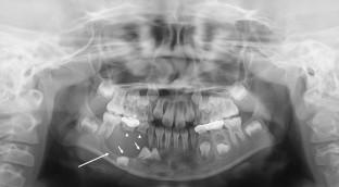
Abstract
Radicular cysts are the most common cystic lesions in the oral cavity, and have a rare occurrence in the primary dentition. We report a case of radicular cyst of mandible in child by multimodal imaging including panoramic radiography, CT, and MR imaging. A 7-year-old girl presented with swelling and without pain, and hypoesthesia on the right side of the mandible. On clinical examination, an expansive lesion with undulation was found to the buccal cortex of the right side of the mandible. Panoramic radiograph showed a unilocular radiolucency with well-defined margin, displaced tooth, and root resorption in the right mandible. Regarding CT imaging, axial soft tissue algorithm CT and bone tissue algorithm CT showed a low-attenuation internal structure and expansion of the buccal cortex of the right side of the mandible. Three-dimensional-CT showed expansion of the buccal cortex of the right side of the mandible. Multiplanar reformation imaging showed displaced tooth, root resorption, and expansion of the buccal cortex of the right side of the mandible. On T1-weighted image, the expansive lesion showed low signal intensity, and T2-weighted and STIR images revealed high signal intensity. A partial biopsy of the mandibular region was performed. Histopathological diagnosis was radicular cyst caused by apical periodontitis with abscess. This case suggests that multimodal imaging, especially CT and MR imaging, could be effective for evaluating mandibular lesions in child.
期刊介绍:
As the official English-language journal of the Japanese Society for Oral and Maxillofacial Radiology and the Asian Academy of Oral and Maxillofacial Radiology, Oral Radiology is intended to be a forum for international collaboration in head and neck diagnostic imaging and all related fields. Oral Radiology features cutting-edge research papers, review articles, case reports, and technical notes from both the clinical and experimental fields. As membership in the Society is not a prerequisite, contributions are welcome from researchers and clinicians worldwide.


 分享
分享
 求助内容:
求助内容: 应助结果提醒方式:
 扫码关注我们
