Zade Moureiden, Hammad Tashkandi, Mohammad Omar Hussaini
{"title":"Sclerotic marginal zone lymphoma: A case report.","authors":"Zade Moureiden, Hammad Tashkandi, Mohammad Omar Hussaini","doi":"10.5662/wjm.v13.i4.366","DOIUrl":null,"url":null,"abstract":"<p><strong>Background: </strong>Marginal zone lymphoma (MZL) is an indolent non-Hodgkin B cell lymphoma with various architectural pattern including perifollicular, follicular colonization, nodular, micronodular, and diffuse patterns. A sclerotic variant has not been previously reported and represents a diagnostic pitfall.</p><p><strong>Case summary: </strong>A 66-year-old male developed left upper extremity swelling. Chest computed tomography (CT) in September 2020 showed 14 cm mass in left axilla. Needle core biopsy of axillary lymph node showed sclerotic tissue with atypical B lymphoid infiltrate but was non-diagnostic. Excisional biopsy was performed for diagnosis and showed extensive fibrosis and minor component of infiltrating B cells. Flow cytometry showed a small population of CD5-, CD10-, kappa restricted B cells. Monoclonal immunoglobulin heavy chain and light chain gene rearrangement were identified. Upon being diagnosed with MZL, patient was treated with rituximab, cyclophosphamide, doxorubicin, vincristine, and prednisone and achieved complete remission by positron emission tomography/CT.</p><p><strong>Conclusion: </strong>This is an important case report because by morphology this case could have easily been overlooked as non-specific fibrosis with chronic inflammation representing a significant diagnostic pitfall. Moreover, this constitutes a new architectural pattern. While sclerotic lymphomas have rarely been described (often misdiagnosed as retroperitoneal fibrosis), we do not know of any cases describing this architectural presentation of MZL.</p>","PeriodicalId":94271,"journal":{"name":"World journal of methodology","volume":"13 4","pages":"366-372"},"PeriodicalIF":0.0000,"publicationDate":"2023-09-20","publicationTypes":"Journal Article","fieldsOfStudy":null,"isOpenAccess":false,"openAccessPdf":"https://ftp.ncbi.nlm.nih.gov/pub/pmc/oa_pdf/d4/96/WJM-13-366.PMC10523246.pdf","citationCount":"0","resultStr":null,"platform":"Semanticscholar","paperid":null,"PeriodicalName":"World journal of methodology","FirstCategoryId":"1085","ListUrlMain":"https://doi.org/10.5662/wjm.v13.i4.366","RegionNum":0,"RegionCategory":null,"ArticlePicture":[],"TitleCN":null,"AbstractTextCN":null,"PMCID":null,"EPubDate":"","PubModel":"","JCR":"","JCRName":"","Score":null,"Total":0}
引用次数: 0
Abstract
Background: Marginal zone lymphoma (MZL) is an indolent non-Hodgkin B cell lymphoma with various architectural pattern including perifollicular, follicular colonization, nodular, micronodular, and diffuse patterns. A sclerotic variant has not been previously reported and represents a diagnostic pitfall.
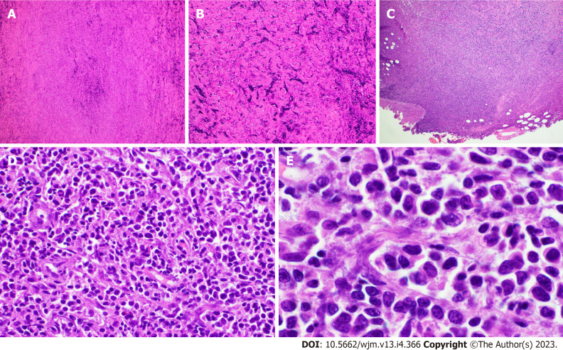
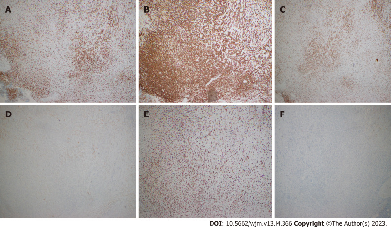
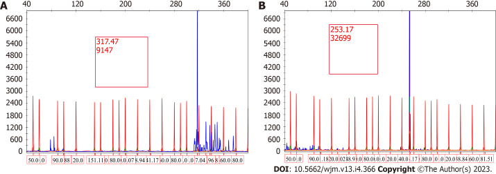
Case summary: A 66-year-old male developed left upper extremity swelling. Chest computed tomography (CT) in September 2020 showed 14 cm mass in left axilla. Needle core biopsy of axillary lymph node showed sclerotic tissue with atypical B lymphoid infiltrate but was non-diagnostic. Excisional biopsy was performed for diagnosis and showed extensive fibrosis and minor component of infiltrating B cells. Flow cytometry showed a small population of CD5-, CD10-, kappa restricted B cells. Monoclonal immunoglobulin heavy chain and light chain gene rearrangement were identified. Upon being diagnosed with MZL, patient was treated with rituximab, cyclophosphamide, doxorubicin, vincristine, and prednisone and achieved complete remission by positron emission tomography/CT.
Conclusion: This is an important case report because by morphology this case could have easily been overlooked as non-specific fibrosis with chronic inflammation representing a significant diagnostic pitfall. Moreover, this constitutes a new architectural pattern. While sclerotic lymphomas have rarely been described (often misdiagnosed as retroperitoneal fibrosis), we do not know of any cases describing this architectural presentation of MZL.


 分享
分享

 求助内容:
求助内容: 应助结果提醒方式:
 扫码关注我们
