{"title":"Dynamics of actomyosin filaments in the contractile ring revealed by ultrastructural analysis","authors":"Takeru Arima, Keisuke Okita, Shigehiko Yumura","doi":"10.1111/gtc.13073","DOIUrl":null,"url":null,"abstract":"<p>Cytokinesis, the final process of cell division, involves the accumulation of actin and myosin II filaments at the cell's equator, forming a contractile ring that facilitates the division into two daughter cells. While light microscopy has provided valuable insights into the molecular mechanism of this process, it has limitations in examining individual filaments in vivo. In this study, we utilized transmission electron microscopy to observe actin and myosin II filaments in the contractile rings of dividing <i>Dictyostelium</i> cells. To synchronize cytokinesis, we developed a novel method that allowed us to visualize dividing cells undergoing cytokinesis with a frequency as high as 18%. This improvement enabled us to examine the lengths and alignments of individual filaments within the contractile rings. As the furrow constricted, the length of actin filaments gradually decreased. Moreover, both actin and myosin II filaments reoriented perpendicularly to the long axis during furrow constriction. Through experiments involving myosin II null cells, we discovered that myosin II plays a role in regulating both the lengths and alignments of actin filaments. Additionally, dynamin-like protein A was found to contribute to regulating the length of actin filaments, while cortexillins were involved in regulating their alignment.</p>","PeriodicalId":12742,"journal":{"name":"Genes to Cells","volume":"28 12","pages":"845-856"},"PeriodicalIF":1.3000,"publicationDate":"2023-10-16","publicationTypes":"Journal Article","fieldsOfStudy":null,"isOpenAccess":false,"openAccessPdf":"","citationCount":"0","resultStr":null,"platform":"Semanticscholar","paperid":null,"PeriodicalName":"Genes to Cells","FirstCategoryId":"99","ListUrlMain":"https://onlinelibrary.wiley.com/doi/10.1111/gtc.13073","RegionNum":4,"RegionCategory":"生物学","ArticlePicture":[],"TitleCN":null,"AbstractTextCN":null,"PMCID":null,"EPubDate":"","PubModel":"","JCR":"Q4","JCRName":"CELL BIOLOGY","Score":null,"Total":0}
引用次数: 0
Abstract
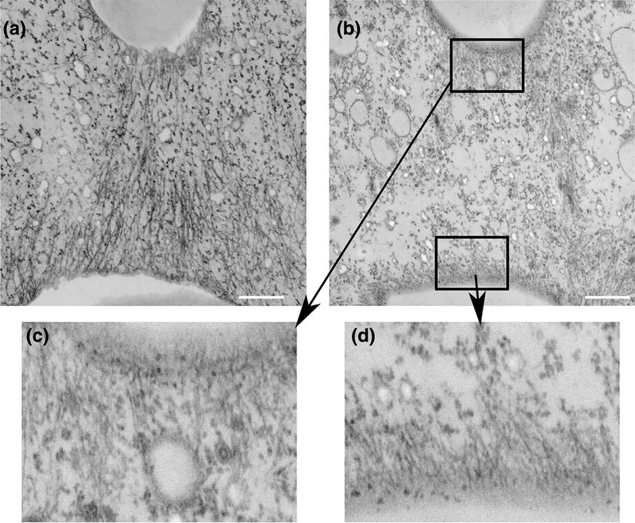
Cytokinesis, the final process of cell division, involves the accumulation of actin and myosin II filaments at the cell's equator, forming a contractile ring that facilitates the division into two daughter cells. While light microscopy has provided valuable insights into the molecular mechanism of this process, it has limitations in examining individual filaments in vivo. In this study, we utilized transmission electron microscopy to observe actin and myosin II filaments in the contractile rings of dividing Dictyostelium cells. To synchronize cytokinesis, we developed a novel method that allowed us to visualize dividing cells undergoing cytokinesis with a frequency as high as 18%. This improvement enabled us to examine the lengths and alignments of individual filaments within the contractile rings. As the furrow constricted, the length of actin filaments gradually decreased. Moreover, both actin and myosin II filaments reoriented perpendicularly to the long axis during furrow constriction. Through experiments involving myosin II null cells, we discovered that myosin II plays a role in regulating both the lengths and alignments of actin filaments. Additionally, dynamin-like protein A was found to contribute to regulating the length of actin filaments, while cortexillins were involved in regulating their alignment.
期刊介绍:
Genes to Cells provides an international forum for the publication of papers describing important aspects of molecular and cellular biology. The journal aims to present papers that provide conceptual advance in the relevant field. Particular emphasis will be placed on work aimed at understanding the basic mechanisms underlying biological events.


 分享
分享
 求助内容:
求助内容: 应助结果提醒方式:
 扫码关注我们
