{"title":"Changes of Beclin-1 and ULK1 in retina of mice model in oxygen-inducedretinopathy","authors":"Jie Wang , Ergang Du , FeiFei Li , Yunliang Zheng","doi":"10.1016/j.aopr.2022.100065","DOIUrl":null,"url":null,"abstract":"<div><h3>Purpose</h3><p>To observe the expression differences and potential effects of autophagy-related Beclin1 (mammalian Atg6) and Uncoordinated-51 like kinase 1 (ULK1) in the oxygen-induced retinopathy (OIR) model.</p></div><div><h3>Materials and methods</h3><p>Thirty-three C57BL/6 mice in OIR model group were exposed to 75 ± 0.5% oxygen from postnatal day-of-life 7 (P7) to P12, and were then brought into normal room environment (relative hypoxia stage) and raised to P17. Thirty-three control mice were kept in a normal room environment. The expression of autophagy in the retina tissue was assessed by Western blot analysis. The thickness and ultrastructural of retina were observed by light microscopy and transmission electron microscope (TEM) on P17.</p></div><div><h3>Results</h3><p>In the hyperoxia stage (P8–P11), the expression of Beclin1, ULK1 and Autophagy 5 (Atg5) in retina showed no significant difference between the OIR model group and the control group. In the relatively hypoxia stage (P14 to P17), however, the protein level of Beclin1, ULK1, and Bcl-2-associated X protein (Bax) were upregulated in the retina of the OIR model group, whereas B-cell lymphoma 2 (Bcl-2) was downregulated. The autophagosomes in the photoreceptors of retina in the OIR mice were observed. The inner-segment/out-segment (IS/OS) layer in OIR model group was thinner than that the control group on P17.</p></div><div><h3>Conclusions</h3><p>The expression of Beclin-1 and ULK1 in retina has changed in the OIR model, and the change of Beclin-1 and ULK1 expression is related to the change of oxygen concentration.</p></div>","PeriodicalId":72103,"journal":{"name":"Advances in ophthalmology practice and research","volume":"2 3","pages":"Article 100065"},"PeriodicalIF":3.4000,"publicationDate":"2022-11-01","publicationTypes":"Journal Article","fieldsOfStudy":null,"isOpenAccess":false,"openAccessPdf":"https://ftp.ncbi.nlm.nih.gov/pub/pmc/oa_pdf/0b/9b/main.PMC10577824.pdf","citationCount":"0","resultStr":null,"platform":"Semanticscholar","paperid":null,"PeriodicalName":"Advances in ophthalmology practice and research","FirstCategoryId":"1085","ListUrlMain":"https://www.sciencedirect.com/science/article/pii/S2667376222000427","RegionNum":0,"RegionCategory":null,"ArticlePicture":[],"TitleCN":null,"AbstractTextCN":null,"PMCID":null,"EPubDate":"","PubModel":"","JCR":"","JCRName":"","Score":null,"Total":0}
引用次数: 0
Abstract
Purpose
To observe the expression differences and potential effects of autophagy-related Beclin1 (mammalian Atg6) and Uncoordinated-51 like kinase 1 (ULK1) in the oxygen-induced retinopathy (OIR) model.
Materials and methods
Thirty-three C57BL/6 mice in OIR model group were exposed to 75 ± 0.5% oxygen from postnatal day-of-life 7 (P7) to P12, and were then brought into normal room environment (relative hypoxia stage) and raised to P17. Thirty-three control mice were kept in a normal room environment. The expression of autophagy in the retina tissue was assessed by Western blot analysis. The thickness and ultrastructural of retina were observed by light microscopy and transmission electron microscope (TEM) on P17.
Results
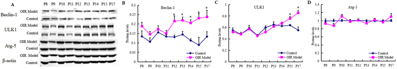
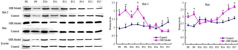
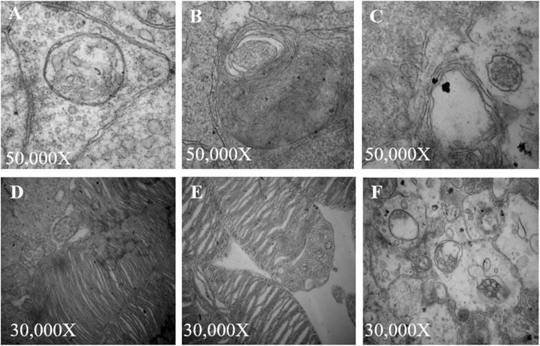
In the hyperoxia stage (P8–P11), the expression of Beclin1, ULK1 and Autophagy 5 (Atg5) in retina showed no significant difference between the OIR model group and the control group. In the relatively hypoxia stage (P14 to P17), however, the protein level of Beclin1, ULK1, and Bcl-2-associated X protein (Bax) were upregulated in the retina of the OIR model group, whereas B-cell lymphoma 2 (Bcl-2) was downregulated. The autophagosomes in the photoreceptors of retina in the OIR mice were observed. The inner-segment/out-segment (IS/OS) layer in OIR model group was thinner than that the control group on P17.
Conclusions
The expression of Beclin-1 and ULK1 in retina has changed in the OIR model, and the change of Beclin-1 and ULK1 expression is related to the change of oxygen concentration.


 分享
分享
 求助内容:
求助内容: 应助结果提醒方式:
 扫码关注我们
