{"title":"Clinical characteristics of radiation-induced optic neuropathy: A single-center retrospective study","authors":"Yongping Wang, Junxia Fu, Huanfen Zhou, Hongen Li, Quangang Xu, Shihui Wei","doi":"10.1016/j.aopr.2023.05.003","DOIUrl":null,"url":null,"abstract":"<div><h3>Purpose</h3><p>To observe the clinical and imaging characteristics of radiation-induced optic neuropathy (RION).</p></div><div><h3>Methods</h3><p>We retrospectively reviewed the clinical data of 43 patients (69 eyes) who were diagnosed with RION at the Chinese PLA General Hospital from 2010 to 2021.</p></div><div><h3>Results</h3><p>The latency from radiotherapy to onset of visual loss ranged from 1 to 132 (36.33 ± 30.48) months. Optic disc pallor and optic disc edema were found in 27.0% (10/37) and 8.1% (3/37) of the eyes, respectively, within 2 months. After treatment, the best corrected visual acuity (BCVA) was restored in 24.6% (17/69) of the eyes and the final BCVA improved in 13.0% (9/69) of the eyes. An 82.5% (33/40) of the eyes with magnetic resonance imaging (MRI) showed enhancement of the affected optic nerve, mostly (69.7%) in the intracranial segment, and 36.4% (12/33) of the eyes with expansion and T2-high signals also showed enhancement of the affected optic nerve. The superior retinal nerve fiber layer (RNFL) and the outer circle superior quadrant (OS) of the inner limiting membrane to retinal pigment epithelium (ILM-RPE) layer thinned significantly during the first month. The center of the ILM-RPE layer thickened significantly during the first two months and the inner circle temporal quadrant (IT) of the ILM-RPE layer thickened significantly from the third to sixth month. The RNFL thinned significantly after 6 months except for the temporal quadrant, and the average inner circle superior quadrant (IS) and outer circle of the ILM-RPE layer thinned significantly after 6 months. There was no significant difference between hyperbaric oxygen therapy (HBOT) and high-dose intravenous methylprednisolone (IVMP) therapy in improving BCVA recovery or final BCVA (<em>P</em> > 0.05).</p></div><div><h3>Conclusions</h3><p>The structural damage of the RNFL and ILM-RPE layer occurred during the first month, the RNFL showed progressive thinning during the follow-up period, while the ILM-RPE layer showed thinning during the first month, thickening from the third to sixth month, and thinning after 6 months. There was a discrete region of enhancement of the optic nerve, often with expansion and high-T2 signals on MRI. HBOT and high-dose IVMP therapy were hardly effective for treating RION in the non-acute stage.</p></div>","PeriodicalId":72103,"journal":{"name":"Advances in ophthalmology practice and research","volume":"3 3","pages":"Pages 141-146"},"PeriodicalIF":3.4000,"publicationDate":"2023-08-01","publicationTypes":"Journal Article","fieldsOfStudy":null,"isOpenAccess":false,"openAccessPdf":"https://ftp.ncbi.nlm.nih.gov/pub/pmc/oa_pdf/ee/be/main.PMC10577828.pdf","citationCount":"0","resultStr":null,"platform":"Semanticscholar","paperid":null,"PeriodicalName":"Advances in ophthalmology practice and research","FirstCategoryId":"1085","ListUrlMain":"https://www.sciencedirect.com/science/article/pii/S2667376223000161","RegionNum":0,"RegionCategory":null,"ArticlePicture":[],"TitleCN":null,"AbstractTextCN":null,"PMCID":null,"EPubDate":"","PubModel":"","JCR":"","JCRName":"","Score":null,"Total":0}
引用次数: 0
Abstract
Purpose
To observe the clinical and imaging characteristics of radiation-induced optic neuropathy (RION).
Methods
We retrospectively reviewed the clinical data of 43 patients (69 eyes) who were diagnosed with RION at the Chinese PLA General Hospital from 2010 to 2021.
Results
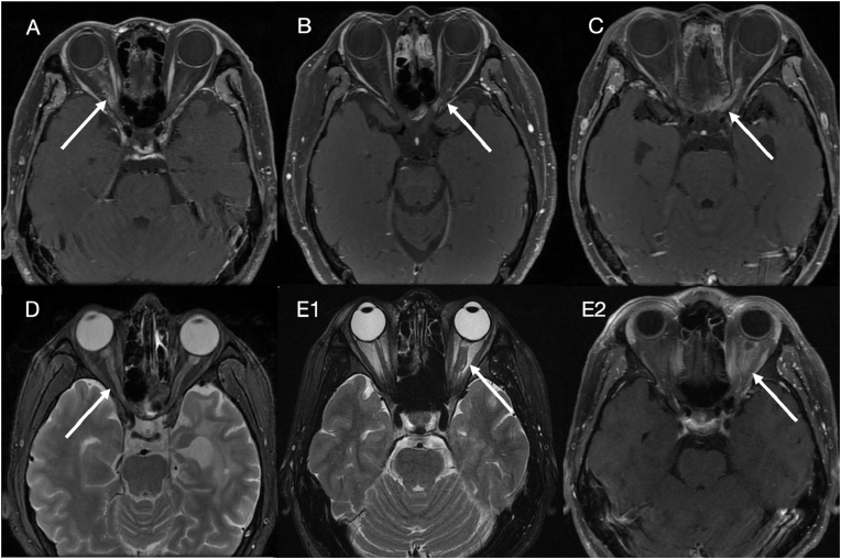
The latency from radiotherapy to onset of visual loss ranged from 1 to 132 (36.33 ± 30.48) months. Optic disc pallor and optic disc edema were found in 27.0% (10/37) and 8.1% (3/37) of the eyes, respectively, within 2 months. After treatment, the best corrected visual acuity (BCVA) was restored in 24.6% (17/69) of the eyes and the final BCVA improved in 13.0% (9/69) of the eyes. An 82.5% (33/40) of the eyes with magnetic resonance imaging (MRI) showed enhancement of the affected optic nerve, mostly (69.7%) in the intracranial segment, and 36.4% (12/33) of the eyes with expansion and T2-high signals also showed enhancement of the affected optic nerve. The superior retinal nerve fiber layer (RNFL) and the outer circle superior quadrant (OS) of the inner limiting membrane to retinal pigment epithelium (ILM-RPE) layer thinned significantly during the first month. The center of the ILM-RPE layer thickened significantly during the first two months and the inner circle temporal quadrant (IT) of the ILM-RPE layer thickened significantly from the third to sixth month. The RNFL thinned significantly after 6 months except for the temporal quadrant, and the average inner circle superior quadrant (IS) and outer circle of the ILM-RPE layer thinned significantly after 6 months. There was no significant difference between hyperbaric oxygen therapy (HBOT) and high-dose intravenous methylprednisolone (IVMP) therapy in improving BCVA recovery or final BCVA (P > 0.05).
Conclusions
The structural damage of the RNFL and ILM-RPE layer occurred during the first month, the RNFL showed progressive thinning during the follow-up period, while the ILM-RPE layer showed thinning during the first month, thickening from the third to sixth month, and thinning after 6 months. There was a discrete region of enhancement of the optic nerve, often with expansion and high-T2 signals on MRI. HBOT and high-dose IVMP therapy were hardly effective for treating RION in the non-acute stage.


 分享
分享
 求助内容:
求助内容: 应助结果提醒方式:
 扫码关注我们
