Xuerui Zhang , Yuan Yang , Yanjun Wen , Haodong Xiao , Jie Peng , Peiquan Zhao
{"title":"Description and surgical management of epiretinal membrane due to combined hamartoma of the retina and retinal pigment epithelium","authors":"Xuerui Zhang , Yuan Yang , Yanjun Wen , Haodong Xiao , Jie Peng , Peiquan Zhao","doi":"10.1016/j.aopr.2022.09.001","DOIUrl":null,"url":null,"abstract":"<div><h3>Purpose</h3><p>To outline the characteristics of Combined Hamartoma of the Retina and Retinal Pigmentation Epithelium (CHRRPE) and provide a comprehensive overview of surgical management of epiretinal membrane (ERM) caused by CHRRPE.</p></div><div><h3>Main text</h3><p>CHRRPE is a rare ocular tumor. It clinically mimics other diseases such as retinoblastoma and choroidal melanoma. The present study reviewed the multimodal imaging of CHRRPE, highlighted the multimodal imaging modalities which are useful for revealing the unique features of CHRRPE and hence allowing physicians to confirm the diagnosis.</p><p>Although most of CHRRPEs are benign harmatoma, progressive visual loss may occur because of the traction of the tumor and other complications. It is treated through surgical removal of the ERM caused by CHRRPE to free retina from the traction. Currently, there is no consensus on the surgical management of CHRRPE. Therefore, the current review was designed to explore the surgical management of ERM caused by CHRRPE and hence provide updated data on this subject.</p></div><div><h3>Conclusions</h3><p>Multimodal imaging technologies, especially optical coherence tomography (OCT), significantly contributes to the diagnosis of CHRRPE and visual prognosis. Surgical management of CHRRPE through removal of ERM is beneficial in patients with worsening VA which is secondary to ERM which is associated with CHRRPE. However, the strategy is limited to patients with long-standing poor vision. However, earlier surgical therapy and subsequent postoperative amblyopia therapy can be explored for children of amblyogenic age.</p></div>","PeriodicalId":72103,"journal":{"name":"Advances in ophthalmology practice and research","volume":"3 1","pages":"Pages 9-14"},"PeriodicalIF":3.4000,"publicationDate":"2023-02-01","publicationTypes":"Journal Article","fieldsOfStudy":null,"isOpenAccess":false,"openAccessPdf":"https://ftp.ncbi.nlm.nih.gov/pub/pmc/oa_pdf/07/91/main.PMC10577870.pdf","citationCount":"0","resultStr":null,"platform":"Semanticscholar","paperid":null,"PeriodicalName":"Advances in ophthalmology practice and research","FirstCategoryId":"1085","ListUrlMain":"https://www.sciencedirect.com/science/article/pii/S2667376222000671","RegionNum":0,"RegionCategory":null,"ArticlePicture":[],"TitleCN":null,"AbstractTextCN":null,"PMCID":null,"EPubDate":"","PubModel":"","JCR":"","JCRName":"","Score":null,"Total":0}
引用次数: 0
Abstract
Purpose
To outline the characteristics of Combined Hamartoma of the Retina and Retinal Pigmentation Epithelium (CHRRPE) and provide a comprehensive overview of surgical management of epiretinal membrane (ERM) caused by CHRRPE.
Main text
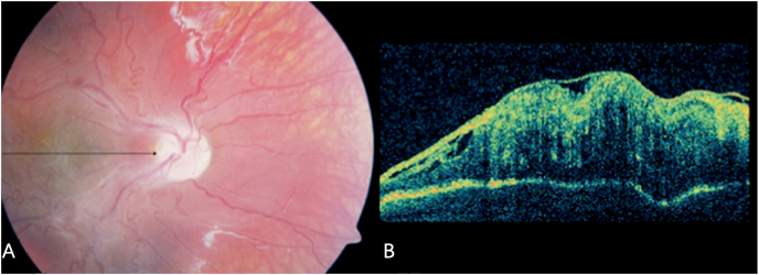
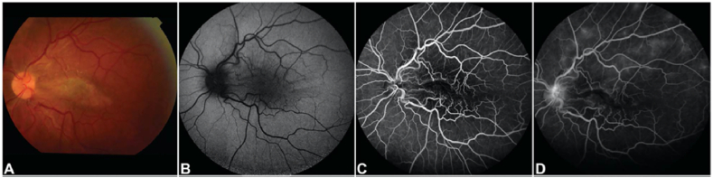
CHRRPE is a rare ocular tumor. It clinically mimics other diseases such as retinoblastoma and choroidal melanoma. The present study reviewed the multimodal imaging of CHRRPE, highlighted the multimodal imaging modalities which are useful for revealing the unique features of CHRRPE and hence allowing physicians to confirm the diagnosis.
Although most of CHRRPEs are benign harmatoma, progressive visual loss may occur because of the traction of the tumor and other complications. It is treated through surgical removal of the ERM caused by CHRRPE to free retina from the traction. Currently, there is no consensus on the surgical management of CHRRPE. Therefore, the current review was designed to explore the surgical management of ERM caused by CHRRPE and hence provide updated data on this subject.
Conclusions
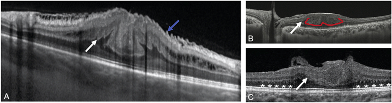
Multimodal imaging technologies, especially optical coherence tomography (OCT), significantly contributes to the diagnosis of CHRRPE and visual prognosis. Surgical management of CHRRPE through removal of ERM is beneficial in patients with worsening VA which is secondary to ERM which is associated with CHRRPE. However, the strategy is limited to patients with long-standing poor vision. However, earlier surgical therapy and subsequent postoperative amblyopia therapy can be explored for children of amblyogenic age.


 分享
分享
 求助内容:
求助内容: 应助结果提醒方式:
 扫码关注我们
