Parthapratim Munshi, Christopher B Stanley, Sudipa Ghimire-Rijal, Xun Lu, Dean A Myles, Matthew J Cuneo
{"title":"Molecular details of ligand selectivity determinants in a promiscuous β-glucan periplasmic binding protein","authors":"Parthapratim Munshi, Christopher B Stanley, Sudipa Ghimire-Rijal, Xun Lu, Dean A Myles, Matthew J Cuneo","doi":"10.1186/1472-6807-13-18","DOIUrl":null,"url":null,"abstract":"<p>Members of the periplasmic binding protein (PBP) superfamily utilize a highly conserved inter-domain ligand binding site that adapts to specifically bind a chemically diverse range of ligands. This paradigm of PBP ligand binding specificity was recently altered when the structure of the <i>Thermotoga maritima</i> cellobiose-binding protein (tmCBP) was solved. The tmCBP binding site is bipartite, comprising a canonical solvent-excluded region (subsite one), adjacent to a solvent-filled cavity (subsite two) where specific and semi-specific ligand recognition occur, respectively.</p><p>A molecular level understanding of binding pocket adaptation mechanisms that simultaneously allow both ligand specificity at subsite one and promiscuity at subsite two has potentially important implications in ligand binding and drug design studies. We sought to investigate the determinants of ligand binding selectivity in tmCBP through biophysical characterization of tmCBP in the presence of varying β-glucan oligosaccharides. Crystal structures show that whilst the amino acids that comprise both the tmCBP subsite one and subsite two binding sites remain fixed in conformation regardless of which ligands are present, the rich hydrogen bonding potential of water molecules may facilitate the ordering and the plasticity of this unique PBP binding site.</p><p>The identification of the roles these water molecules play in ligand recognition suggests potential mechanisms that can be utilized to adapt a single ligand binding site to recognize multiple distinct ligands.</p>","PeriodicalId":498,"journal":{"name":"BMC Structural Biology","volume":"13 1","pages":""},"PeriodicalIF":2.2220,"publicationDate":"2013-10-04","publicationTypes":"Journal Article","fieldsOfStudy":null,"isOpenAccess":false,"openAccessPdf":"https://sci-hub-pdf.com/10.1186/1472-6807-13-18","citationCount":"7","resultStr":null,"platform":"Semanticscholar","paperid":null,"PeriodicalName":"BMC Structural Biology","FirstCategoryId":"1085","ListUrlMain":"https://link.springer.com/article/10.1186/1472-6807-13-18","RegionNum":0,"RegionCategory":null,"ArticlePicture":[],"TitleCN":null,"AbstractTextCN":null,"PMCID":null,"EPubDate":"","PubModel":"","JCR":"Q3","JCRName":"Biochemistry, Genetics and Molecular Biology","Score":null,"Total":0}
引用次数: 7
Abstract
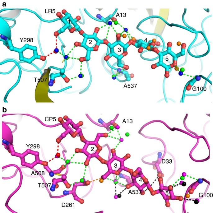
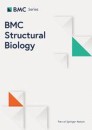
Members of the periplasmic binding protein (PBP) superfamily utilize a highly conserved inter-domain ligand binding site that adapts to specifically bind a chemically diverse range of ligands. This paradigm of PBP ligand binding specificity was recently altered when the structure of the Thermotoga maritima cellobiose-binding protein (tmCBP) was solved. The tmCBP binding site is bipartite, comprising a canonical solvent-excluded region (subsite one), adjacent to a solvent-filled cavity (subsite two) where specific and semi-specific ligand recognition occur, respectively.
A molecular level understanding of binding pocket adaptation mechanisms that simultaneously allow both ligand specificity at subsite one and promiscuity at subsite two has potentially important implications in ligand binding and drug design studies. We sought to investigate the determinants of ligand binding selectivity in tmCBP through biophysical characterization of tmCBP in the presence of varying β-glucan oligosaccharides. Crystal structures show that whilst the amino acids that comprise both the tmCBP subsite one and subsite two binding sites remain fixed in conformation regardless of which ligands are present, the rich hydrogen bonding potential of water molecules may facilitate the ordering and the plasticity of this unique PBP binding site.
The identification of the roles these water molecules play in ligand recognition suggests potential mechanisms that can be utilized to adapt a single ligand binding site to recognize multiple distinct ligands.
期刊介绍:
BMC Structural Biology is an open access, peer-reviewed journal that considers articles on investigations into the structure of biological macromolecules, including solving structures, structural and functional analyses, and computational modeling.


 分享
分享
 求助内容:
求助内容: 应助结果提醒方式:
 扫码关注我们
