Kamal L Nahas, João Ferreira Fernandes, Nina Vyas, Colin Crump, Stephen Graham, Maria Harkiolaki
{"title":"<i>Contour</i>, a semi-automated segmentation and quantitation tool for cryo-soft-X-ray tomography.","authors":"Kamal L Nahas, João Ferreira Fernandes, Nina Vyas, Colin Crump, Stephen Graham, Maria Harkiolaki","doi":"10.1017/S2633903X22000046","DOIUrl":null,"url":null,"abstract":"<p><p>Cryo-soft-X-ray tomography is being increasingly used in biological research to study the morphology of cellular compartments and how they change in response to different stimuli, such as viral infections. Segmentation of these compartments is limited by time-consuming manual tools or machine learning algorithms that require extensive time and effort to train. Here we describe <i>Contour</i>, a new, easy-to-use, highly automated segmentation tool that enables accelerated segmentation of tomograms to delineate distinct cellular compartments. Using <i>Contour</i>, cellular structures can be segmented based on their projection intensity and geometrical width by applying a threshold range to the image and excluding noise smaller in width than the cellular compartments of interest. This method is less laborious and less prone to errors from human judgement than current tools that require features to be manually traced, and does not require training datasets as would machine-learning driven segmentation. We show that high-contrast compartments such as mitochondria, lipid droplets, and features at the cell surface can be easily segmented with this technique in the context of investigating herpes simplex virus 1 infection. <i>Contour</i> can extract geometric measurements from 3D segmented volumes, providing a new method to quantitate cryo-soft-X-ray tomography data. <i>Contour</i> can be freely downloaded at github.com/kamallouisnahas/Contour.</p>","PeriodicalId":72371,"journal":{"name":"Biological imaging","volume":"2 1","pages":""},"PeriodicalIF":0.0000,"publicationDate":"2022-05-17","publicationTypes":"Journal Article","fieldsOfStudy":null,"isOpenAccess":false,"openAccessPdf":"https://www.ncbi.nlm.nih.gov/pmc/articles/PMC7612748/pdf/","citationCount":"0","resultStr":null,"platform":"Semanticscholar","paperid":null,"PeriodicalName":"Biological imaging","FirstCategoryId":"1085","ListUrlMain":"https://doi.org/10.1017/S2633903X22000046","RegionNum":0,"RegionCategory":null,"ArticlePicture":[],"TitleCN":null,"AbstractTextCN":null,"PMCID":null,"EPubDate":"2022/1/1 0:00:00","PubModel":"eCollection","JCR":"","JCRName":"","Score":null,"Total":0}
引用次数: 0
Abstract
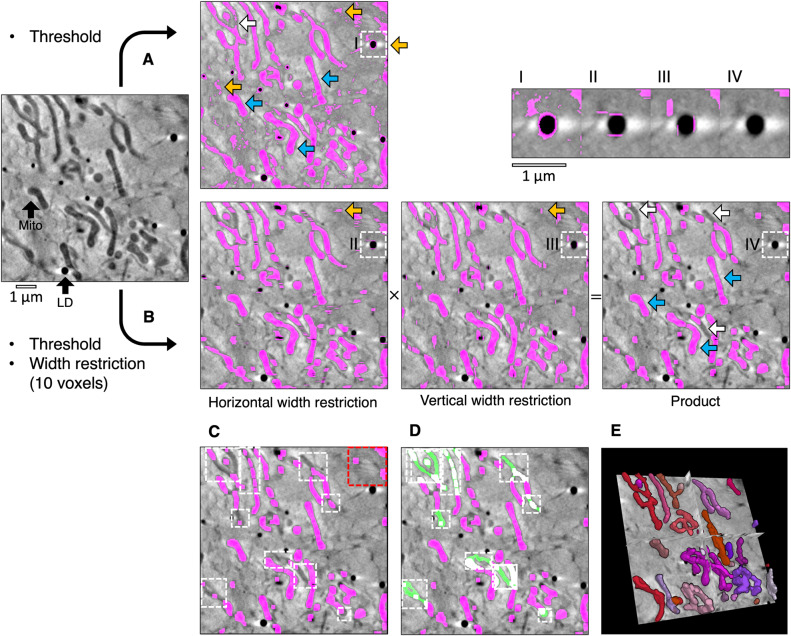
Cryo-soft-X-ray tomography is being increasingly used in biological research to study the morphology of cellular compartments and how they change in response to different stimuli, such as viral infections. Segmentation of these compartments is limited by time-consuming manual tools or machine learning algorithms that require extensive time and effort to train. Here we describe Contour, a new, easy-to-use, highly automated segmentation tool that enables accelerated segmentation of tomograms to delineate distinct cellular compartments. Using Contour, cellular structures can be segmented based on their projection intensity and geometrical width by applying a threshold range to the image and excluding noise smaller in width than the cellular compartments of interest. This method is less laborious and less prone to errors from human judgement than current tools that require features to be manually traced, and does not require training datasets as would machine-learning driven segmentation. We show that high-contrast compartments such as mitochondria, lipid droplets, and features at the cell surface can be easily segmented with this technique in the context of investigating herpes simplex virus 1 infection. Contour can extract geometric measurements from 3D segmented volumes, providing a new method to quantitate cryo-soft-X-ray tomography data. Contour can be freely downloaded at github.com/kamallouisnahas/Contour.


 分享
分享

 求助内容:
求助内容: 应助结果提醒方式:
 扫码关注我们
