Chemotherapy, particularly oxaliplatin, has been associated with the development of focal nodular hyperplasia (FNH). Imaging diagnosis of FNH is well standardized, but it can be misdiagnosed as liver metastasis. The aim of this study was to describe the pathological features of FNH occurring after systemic chemotherapy.
From our pathological files for 1990-2021, we retrieved 15 cases of resected newly developed FNH in adults with liver metastasis treated with systemic chemotherapy. Pathological features of FNH nodules and non-tumoral liver samples were reviewed.
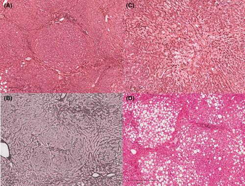
In 11/15 (73%) cases, FNH developed after an oxaliplatin-based regimen. The median interval from the beginning of chemotherapy to the FNH diagnosis was 15 months. FNH was unique in 11 (73%) cases, and the median size of nodules was 1.1 cm [range 0.5-2.5]. Histologically, 9 (60%), 11 (73%) and 11 (73%) cases exhibited fibrous central scar, dystrophic vessels and ductular proliferation, respectively, with all three criteria present in five (33%) cases. Eight (53%) cases showed intralesional steatosis and nine (60%) cases showed a glutamine synthetase immunostaining map-like pattern. In non-tumoral liver, eight (53%) cases exhibited sinusoidal obstruction syndrome and four (27%) nodular regenerative hyperplasia.
The occurrence of FNH after systemic chemotherapy is an emerging condition challenging the imaging diagnosis because typical morphological features are frequently missing. The presence of sinusoidal changes, including regenerative hyperplasia, in non-tumoral liver supports the potential role of chemotherapy in the pathogenesis of FNH.


