Sarin Chimnaronk, Jatuporn Sitthiroongruang, Kanokporn Srisucharitpanit, Monrudee Srisaisup, Albert J. Ketterman, Panadda Boonserm
{"title":"The crystal structure of JNK from Drosophila melanogaster reveals an evolutionarily conserved topology with that of mammalian JNK proteins","authors":"Sarin Chimnaronk, Jatuporn Sitthiroongruang, Kanokporn Srisucharitpanit, Monrudee Srisaisup, Albert J. Ketterman, Panadda Boonserm","doi":"10.1186/s12900-015-0045-1","DOIUrl":null,"url":null,"abstract":"<p>The c-Jun N-terminal kinases (JNKs), members of the mitogen-activated protein kinase (MAPK) family, engage in diverse cellular responses to signals produced under normal development and stress conditions. In <i>Drosophila</i>, only one JNK member is present, whereas ten isoforms from three JNK genes (JNK1, 2, and 3) are present in mammalian cells. To date, several mammalian JNK structures have been determined, however, there has been no report of any insect JNK structure.</p><p>We report the first structure of JNK from <i>Drosophila melanogaster</i> (DJNK). The crystal structure of the unphosphorylated form of DJNK complexed with adenylyl imidodiphosphate (AMP-PNP) has been solved at 1.79?? resolution. The fold and topology of DJNK are similar to those of mammalian JNK isoforms, demonstrating their evolutionarily conserved structures and functions. Structural comparisons of DJNK and the closely related mammalian JNKs also allow identification of putative catalytic residues, substrate-binding sites and conformational alterations upon docking interaction with <i>Drosophila</i> scaffold proteins.</p><p>The DJNK structure reveals common features with those of the mammalian JNK isoforms, thereby allowing the mapping of putative catalytic and substrate binding sites. Additionally, structural changes upon peptide binding could be predicted based on the comparison with the closely-related JNK3 structure in complex with pepJIP1. This is the first structure of insect JNK reported to date, and will provide a platform for future mutational studies in <i>Drosophila</i> to ascertain the functional role of insect JNK.</p>","PeriodicalId":498,"journal":{"name":"BMC Structural Biology","volume":"15 1","pages":""},"PeriodicalIF":2.2220,"publicationDate":"2015-09-16","publicationTypes":"Journal Article","fieldsOfStudy":null,"isOpenAccess":false,"openAccessPdf":"https://sci-hub-pdf.com/10.1186/s12900-015-0045-1","citationCount":"11","resultStr":null,"platform":"Semanticscholar","paperid":null,"PeriodicalName":"BMC Structural Biology","FirstCategoryId":"1085","ListUrlMain":"https://link.springer.com/article/10.1186/s12900-015-0045-1","RegionNum":0,"RegionCategory":null,"ArticlePicture":[],"TitleCN":null,"AbstractTextCN":null,"PMCID":null,"EPubDate":"","PubModel":"","JCR":"Q3","JCRName":"Biochemistry, Genetics and Molecular Biology","Score":null,"Total":0}
引用次数: 11
Abstract
The c-Jun N-terminal kinases (JNKs), members of the mitogen-activated protein kinase (MAPK) family, engage in diverse cellular responses to signals produced under normal development and stress conditions. In Drosophila, only one JNK member is present, whereas ten isoforms from three JNK genes (JNK1, 2, and 3) are present in mammalian cells. To date, several mammalian JNK structures have been determined, however, there has been no report of any insect JNK structure.
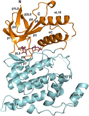
We report the first structure of JNK from Drosophila melanogaster (DJNK). The crystal structure of the unphosphorylated form of DJNK complexed with adenylyl imidodiphosphate (AMP-PNP) has been solved at 1.79?? resolution. The fold and topology of DJNK are similar to those of mammalian JNK isoforms, demonstrating their evolutionarily conserved structures and functions. Structural comparisons of DJNK and the closely related mammalian JNKs also allow identification of putative catalytic residues, substrate-binding sites and conformational alterations upon docking interaction with Drosophila scaffold proteins.
The DJNK structure reveals common features with those of the mammalian JNK isoforms, thereby allowing the mapping of putative catalytic and substrate binding sites. Additionally, structural changes upon peptide binding could be predicted based on the comparison with the closely-related JNK3 structure in complex with pepJIP1. This is the first structure of insect JNK reported to date, and will provide a platform for future mutational studies in Drosophila to ascertain the functional role of insect JNK.
期刊介绍:
BMC Structural Biology is an open access, peer-reviewed journal that considers articles on investigations into the structure of biological macromolecules, including solving structures, structural and functional analyses, and computational modeling.


 分享
分享
 求助内容:
求助内容: 应助结果提醒方式:
 扫码关注我们
