Computed tomography, not bioelectrical impedance analysis, is the proper method for evaluating changes in skeletal muscle mass in liver disease

Masatsugu Ohara, Goki Suda, Megumi Kimura, Osamu Maehara, Tomoe Shimazaki, Taku Shigesawa, Kazuharu Suzuki, Akihisa Nakamura, Naoki Kawagishi, Masato Nakai, Takuya Sho, Mitsuteru Natsuizaka, Kenichi Morikawa, Koji Ogawa, Naoya Sakamoto
{"title":"Computed tomography, not bioelectrical impedance analysis, is the proper method for evaluating changes in skeletal muscle mass in liver disease","authors":"Masatsugu Ohara,&nbsp;Goki Suda,&nbsp;Megumi Kimura,&nbsp;Osamu Maehara,&nbsp;Tomoe Shimazaki,&nbsp;Taku Shigesawa,&nbsp;Kazuharu Suzuki,&nbsp;Akihisa Nakamura,&nbsp;Naoki Kawagishi,&nbsp;Masato Nakai,&nbsp;Takuya Sho,&nbsp;Mitsuteru Natsuizaka,&nbsp;Kenichi Morikawa,&nbsp;Koji Ogawa,&nbsp;Naoya Sakamoto","doi":"10.1002/rco2.20","DOIUrl":null,"url":null,"abstract":"<div>\n \n \n <section>\n \n <h3> Background</h3>\n \n <p>Sarcopenia is associated with poor prognosis in patients with chronic liver disease (CLD). As rapid skeletal muscle wasting predicts worse prognosis and a novel therapy for sarcopenia needs to be evaluated for validation, accurate evaluation methods for relative changes in muscle mass are crucial.</p>\n </section>\n \n <section>\n \n <h3> Methods</h3>\n \n <p>We screened CLD patients who had skeletal muscle mass evaluation between June 2015 and December 2017. Patients were included if they had adequate information, were followed for &gt;6 months, and had skeletal muscle mass evaluation by both bioelectrical impedance analysis (BIA) and computed tomography (CT) imaging at baseline and the second evaluation point. We compared BIA and CT imaging in terms of their ability to quantify skeletal muscle mass and identify relative changes in muscle mass in CLD patients.</p>\n </section>\n \n <section>\n \n <h3> Results</h3>\n \n <p>Of the screened 447 CLD patients, 110 were included in this study, and 71 (64.5%) were men. The median age was 68 (range 21 to 90) years. In total, 83 (75.5%) and 32 (29.1%) patients had liver cirrhosis and hepatocellular carcinoma, respectively. Of them, 50 (45.5%) patients were liver cirrhosis patients without hepatocellular carcinoma through the observation period. Skeletal muscle mass index (SMI) by BIA, psoas muscle mass index (PMI), and SMI based on CT imaging were significantly correlated at baseline [SMI by simple CT method and SMI by BIA (<i>r</i> = 0.61, <i>P</i> &lt; 0.01), SMI by BIA and PMI (<i>r</i> = 0.65, <i>P</i> &lt; 0.01), and SMI by simple CT method and PMI (<i>r</i> = 0.82, <i>P</i> &lt; 0.01), respectively] and second evaluation point [SMI by simple CT method and SMI by BIA (<i>r</i> = 0.51, <i>P</i> &lt; 0.01), SMI by BIA and PMI (<i>r</i> = 0.58, <i>P</i> &lt; 0.01), and SMI by simple CT method and PMI (<i>r</i> = 0.92, <i>P</i> &lt; 0.01), respectively]. Similar to previous reports, based on the PMI and SMI by simple CT method, patients with more severe liver dysfunction experienced more rapid skeletal muscle mass loss (ΔSimple method/years and ΔPMI/years in patients with Child‑Pugh Classes A, B, and C: Child‑Pugh A, −3.34%; B, −11.77%; C, −18.78%; and Child‑Pugh A, −0.78%; B, −6.33%; C, −7.71%, respectively). Completely opposite results were obtained based on SMI by BIA (Child‑Pugh A, −0.70%; B, 1.42%; C, 12.48%). A subgroup analysis revealed that in patients with fluid retention and diuretic administration, SMI by BIA increased with time (<i>P</i> &lt; 0.01).</p>\n </section>\n \n <section>\n \n <h3> Conclusions</h3>\n \n <p>For accurate evaluation of the relative changes in skeletal muscle mass in patients with CLD, CT imaging method, and not BIA, is one of the proper methods.</p>\n </section>\n </div>","PeriodicalId":73544,"journal":{"name":"JCSM rapid communications","volume":null,"pages":null},"PeriodicalIF":0.0000,"publicationDate":"2020-07-15","publicationTypes":"Journal Article","fieldsOfStudy":null,"isOpenAccess":false,"openAccessPdf":"https://sci-hub-pdf.com/10.1002/rco2.20","citationCount":"7","resultStr":null,"platform":"Semanticscholar","paperid":null,"PeriodicalName":"JCSM rapid communications","FirstCategoryId":"1085","ListUrlMain":"https://onlinelibrary.wiley.com/doi/10.1002/rco2.20","RegionNum":0,"RegionCategory":null,"ArticlePicture":[],"TitleCN":null,"AbstractTextCN":null,"PMCID":null,"EPubDate":"","PubModel":"","JCR":"","JCRName":"","Score":null,"Total":0}
引用次数: 7

Abstract

Background

Sarcopenia is associated with poor prognosis in patients with chronic liver disease (CLD). As rapid skeletal muscle wasting predicts worse prognosis and a novel therapy for sarcopenia needs to be evaluated for validation, accurate evaluation methods for relative changes in muscle mass are crucial.

Methods

We screened CLD patients who had skeletal muscle mass evaluation between June 2015 and December 2017. Patients were included if they had adequate information, were followed for >6 months, and had skeletal muscle mass evaluation by both bioelectrical impedance analysis (BIA) and computed tomography (CT) imaging at baseline and the second evaluation point. We compared BIA and CT imaging in terms of their ability to quantify skeletal muscle mass and identify relative changes in muscle mass in CLD patients.

Results

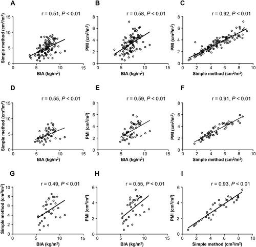
Of the screened 447 CLD patients, 110 were included in this study, and 71 (64.5%) were men. The median age was 68 (range 21 to 90) years. In total, 83 (75.5%) and 32 (29.1%) patients had liver cirrhosis and hepatocellular carcinoma, respectively. Of them, 50 (45.5%) patients were liver cirrhosis patients without hepatocellular carcinoma through the observation period. Skeletal muscle mass index (SMI) by BIA, psoas muscle mass index (PMI), and SMI based on CT imaging were significantly correlated at baseline [SMI by simple CT method and SMI by BIA (r = 0.61, P < 0.01), SMI by BIA and PMI (r = 0.65, P < 0.01), and SMI by simple CT method and PMI (r = 0.82, P < 0.01), respectively] and second evaluation point [SMI by simple CT method and SMI by BIA (r = 0.51, P < 0.01), SMI by BIA and PMI (r = 0.58, P < 0.01), and SMI by simple CT method and PMI (r = 0.92, P < 0.01), respectively]. Similar to previous reports, based on the PMI and SMI by simple CT method, patients with more severe liver dysfunction experienced more rapid skeletal muscle mass loss (ΔSimple method/years and ΔPMI/years in patients with Child‑Pugh Classes A, B, and C: Child‑Pugh A, −3.34%; B, −11.77%; C, −18.78%; and Child‑Pugh A, −0.78%; B, −6.33%; C, −7.71%, respectively). Completely opposite results were obtained based on SMI by BIA (Child‑Pugh A, −0.70%; B, 1.42%; C, 12.48%). A subgroup analysis revealed that in patients with fluid retention and diuretic administration, SMI by BIA increased with time (P < 0.01).

Conclusions

For accurate evaluation of the relative changes in skeletal muscle mass in patients with CLD, CT imaging method, and not BIA, is one of the proper methods.
Abstract Image

查看原文
分享 分享
微信好友 朋友圈 QQ好友 复制链接
本刊更多论文
计算机断层扫描,而不是生物电阻抗分析,是评估肝病骨骼肌质量变化的正确方法
慢性肝病(CLD)患者的Sarcopenia与预后不良有关。由于骨骼肌快速萎缩预示着更糟糕的预后,并且需要评估一种新的少肌症治疗方法进行验证,因此准确评估肌肉质量相对变化的方法至关重要。
本文章由计算机程序翻译,如有差异,请以英文原文为准。
求助全文
约1分钟内获得全文 去求助
来源期刊
自引率
0.00%
发文量
0
审稿时长
10 weeks
期刊最新文献
Issue Information Altered skeletal muscle density: a matter of increased fat infiltration or reduced muscle mass? Uncorrected and subcutaneous fat-corrected echo intensities are similarly associated with magnetic resonance imaging per cent fat A genome wide association study to identify germline copy number variants associated with cancer cachexia: a preliminary analysis Combined orchiectomy and limb immobilization recapitulate early age-related changes to skeletal muscle in mice
×
引用
GB/T 7714-2015
复制
MLA
复制
APA
复制
导出至
BibTeX EndNote RefMan NoteFirst NoteExpress
×
×
提示
您的信息不完整,为了账户安全,请先补充。
现在去补充
×
提示
您因"违规操作"
具体请查看互助需知
我知道了
×
提示
现在去查看 取消
×
提示
确定
0
微信
客服QQ
Book学术公众号 扫码关注我们
反馈
×
意见反馈
请填写您的意见或建议
请填写您的手机或邮箱
已复制链接
已复制链接
快去分享给好友吧!
我知道了
×
扫码分享
扫码分享
Book学术官方微信
Book学术文献互助
Book学术文献互助群
群 号:481959085
Book学术
文献互助 智能选刊 最新文献 互助须知 联系我们:info@booksci.cn
Book学术提供免费学术资源搜索服务,方便国内外学者检索中英文文献。致力于提供最便捷和优质的服务体验。
Copyright © 2023 Book学术 All rights reserved.
ghs 京公网安备 11010802042870号 京ICP备2023020795号-1