Wieke Haakma , Martijn Froeling , Michael Pedersen , Lars Uhrenholt , Perla Douven , Alexander Leemans , Lene Warner Thorup Boel
{"title":"Post-mortem diffusion MRI of the cervical spine and its nerve roots","authors":"Wieke Haakma , Martijn Froeling , Michael Pedersen , Lars Uhrenholt , Perla Douven , Alexander Leemans , Lene Warner Thorup Boel","doi":"10.1016/j.jofri.2018.02.006","DOIUrl":null,"url":null,"abstract":"<div><h3>Purpose</h3><p>The aim of this work is to examine the architectural configuration and the microstructural substrate of the cervical spine and its nerve roots with post-mortem (PM) diffusion tensor imaging (DTI) in non-fixed subjects and to compare these findings with histology.</p></div><div><h3>Methods</h3><p>Magnetic resonance imaging (MRI) data were acquired on a 1.5 T MRI scanner in five non-fixed non-trauma deaths. Two different areas were evaluated: 1) <em>the cervical spinal cord and ventral and dorsal nerve roots</em> with a “high in-plane” DTI and a multi-echo fast field echo protocol, and 2) <span><em>the cervical </em><em>peripheral nerves</em></span> with an “isotropic” DTI and a 3D turbo spin echo protocol. Histology samples were obtained matching the anatomical level of the slices of the ‘high in-plane’ DTI protocol.</p></div><div><h3>Results</h3><p>We were able to show detailed reconstructions of the dorsal and ventral nerve roots with the ‘high in-plane’ protocol and identified a low fractional anisotropy<span> (FA = 0.30 ± 0.08) in the grey matter and a high FA (0.51 ± 0.13) in the white matter. Both grey and white matter configurations correlated with the anatomical MRI, the diffusion MRI, and with the histological sections. Using the ‘isotropic’ DTI protocol, it was feasible to reconstruct the spinal cord, cervical nerves, and nerve roots in all PM subjects.</span></p></div><div><h3>Conclusion</h3><p>We were able to generate detailed architectural configurations of the ventral and dorsal nerve roots. Anatomical and diffusion MR scans showed good qualitative agreement with histology. We believe that PMDTI will be helpful in the assessment of head and neck injuries in a forensic setting.</p></div>","PeriodicalId":45371,"journal":{"name":"Journal of Forensic Radiology and Imaging","volume":"12 ","pages":"Pages 50-56"},"PeriodicalIF":0.0000,"publicationDate":"2018-03-01","publicationTypes":"Journal Article","fieldsOfStudy":null,"isOpenAccess":false,"openAccessPdf":"https://sci-hub-pdf.com/10.1016/j.jofri.2018.02.006","citationCount":"3","resultStr":null,"platform":"Semanticscholar","paperid":null,"PeriodicalName":"Journal of Forensic Radiology and Imaging","FirstCategoryId":"1085","ListUrlMain":"https://www.sciencedirect.com/science/article/pii/S2212478017300801","RegionNum":0,"RegionCategory":null,"ArticlePicture":[],"TitleCN":null,"AbstractTextCN":null,"PMCID":null,"EPubDate":"","PubModel":"","JCR":"","JCRName":"","Score":null,"Total":0}
引用次数: 3
Abstract
Purpose
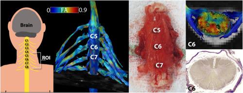
The aim of this work is to examine the architectural configuration and the microstructural substrate of the cervical spine and its nerve roots with post-mortem (PM) diffusion tensor imaging (DTI) in non-fixed subjects and to compare these findings with histology.
Methods
Magnetic resonance imaging (MRI) data were acquired on a 1.5 T MRI scanner in five non-fixed non-trauma deaths. Two different areas were evaluated: 1) the cervical spinal cord and ventral and dorsal nerve roots with a “high in-plane” DTI and a multi-echo fast field echo protocol, and 2) the cervical peripheral nerves with an “isotropic” DTI and a 3D turbo spin echo protocol. Histology samples were obtained matching the anatomical level of the slices of the ‘high in-plane’ DTI protocol.
Results
We were able to show detailed reconstructions of the dorsal and ventral nerve roots with the ‘high in-plane’ protocol and identified a low fractional anisotropy (FA = 0.30 ± 0.08) in the grey matter and a high FA (0.51 ± 0.13) in the white matter. Both grey and white matter configurations correlated with the anatomical MRI, the diffusion MRI, and with the histological sections. Using the ‘isotropic’ DTI protocol, it was feasible to reconstruct the spinal cord, cervical nerves, and nerve roots in all PM subjects.
Conclusion
We were able to generate detailed architectural configurations of the ventral and dorsal nerve roots. Anatomical and diffusion MR scans showed good qualitative agreement with histology. We believe that PMDTI will be helpful in the assessment of head and neck injuries in a forensic setting.


 分享
分享
 求助内容:
求助内容: 应助结果提醒方式:
 扫码关注我们
