Udupi A Ramagopal, Natalya G Dulyaninova, Kristen M Varney, Paul T Wilder, Sridevi Nallamsetty, Michael Brenowitz, David J Weber, Steven C Almo, Anne R Bresnick
{"title":"Structure of the S100A4/myosin-IIA complex","authors":"Udupi A Ramagopal, Natalya G Dulyaninova, Kristen M Varney, Paul T Wilder, Sridevi Nallamsetty, Michael Brenowitz, David J Weber, Steven C Almo, Anne R Bresnick","doi":"10.1186/1472-6807-13-31","DOIUrl":null,"url":null,"abstract":"<p>S100A4, a member of the S100 family of Ca<sup>2+</sup>-binding proteins, modulates the motility of both non-transformed and cancer cells by regulating the localization and stability of cellular protrusions. Biochemical studies have demonstrated that S100A4 binds to the C-terminal end of the myosin-IIA heavy chain coiled-coil and disassembles myosin-IIA filaments; however, the mechanism by which S100A4 mediates myosin-IIA depolymerization is not well understood.</p><p>We determined the X-ray crystal structure of the S100A4Δ8C/MIIA<sup>1908-1923</sup> peptide complex, which showed an asymmetric binding mode for the myosin-IIA peptide across the S100A4 dimer interface. This asymmetric binding mode was confirmed in NMR studies using a spin-labeled myosin-IIA peptide. In addition, our NMR data indicate that S100A4Δ8C binds the MIIA<sup>1908-1923</sup> peptide in an orientation very similar to that observed for wild-type S100A4. Studies of complex formation using a longer, dimeric myosin-IIA construct demonstrated that S100A4 binding dissociates the two myosin-IIA polypeptide chains to form a complex composed of one S100A4 dimer and a single myosin-IIA polypeptide chain. This interaction is mediated, in part, by the instability of the region of the myosin-IIA coiled-coil encompassing the S100A4 binding site.</p><p>The structure of the S100A4/MIIA<sup>1908-1923</sup> peptide complex has revealed the overall architecture of this assembly and the detailed atomic interactions that mediate S100A4 binding to the myosin-IIA heavy chain. These structural studies support the idea that residues 1908–1923 of the myosin-IIA heavy chain represent a core sequence for the S100A4/myosin-IIA complex. In addition, biophysical studies suggest that structural fluctuations within the myosin-IIA coiled-coil may facilitate S100A4 docking onto a single myosin-IIA polypeptide chain.</p>","PeriodicalId":498,"journal":{"name":"BMC Structural Biology","volume":"13 1","pages":""},"PeriodicalIF":2.2220,"publicationDate":"2013-11-20","publicationTypes":"Journal Article","fieldsOfStudy":null,"isOpenAccess":false,"openAccessPdf":"https://sci-hub-pdf.com/10.1186/1472-6807-13-31","citationCount":"21","resultStr":null,"platform":"Semanticscholar","paperid":null,"PeriodicalName":"BMC Structural Biology","FirstCategoryId":"1085","ListUrlMain":"https://link.springer.com/article/10.1186/1472-6807-13-31","RegionNum":0,"RegionCategory":null,"ArticlePicture":[],"TitleCN":null,"AbstractTextCN":null,"PMCID":null,"EPubDate":"","PubModel":"","JCR":"Q3","JCRName":"Biochemistry, Genetics and Molecular Biology","Score":null,"Total":0}
引用次数: 21
Abstract
S100A4, a member of the S100 family of Ca2+-binding proteins, modulates the motility of both non-transformed and cancer cells by regulating the localization and stability of cellular protrusions. Biochemical studies have demonstrated that S100A4 binds to the C-terminal end of the myosin-IIA heavy chain coiled-coil and disassembles myosin-IIA filaments; however, the mechanism by which S100A4 mediates myosin-IIA depolymerization is not well understood.
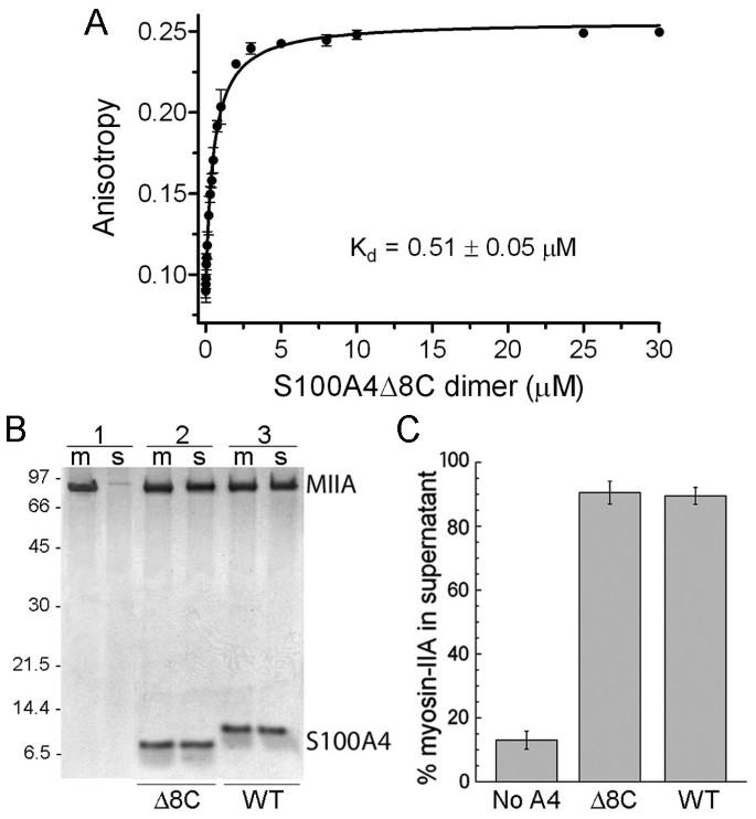
We determined the X-ray crystal structure of the S100A4Δ8C/MIIA1908-1923 peptide complex, which showed an asymmetric binding mode for the myosin-IIA peptide across the S100A4 dimer interface. This asymmetric binding mode was confirmed in NMR studies using a spin-labeled myosin-IIA peptide. In addition, our NMR data indicate that S100A4Δ8C binds the MIIA1908-1923 peptide in an orientation very similar to that observed for wild-type S100A4. Studies of complex formation using a longer, dimeric myosin-IIA construct demonstrated that S100A4 binding dissociates the two myosin-IIA polypeptide chains to form a complex composed of one S100A4 dimer and a single myosin-IIA polypeptide chain. This interaction is mediated, in part, by the instability of the region of the myosin-IIA coiled-coil encompassing the S100A4 binding site.
The structure of the S100A4/MIIA1908-1923 peptide complex has revealed the overall architecture of this assembly and the detailed atomic interactions that mediate S100A4 binding to the myosin-IIA heavy chain. These structural studies support the idea that residues 1908–1923 of the myosin-IIA heavy chain represent a core sequence for the S100A4/myosin-IIA complex. In addition, biophysical studies suggest that structural fluctuations within the myosin-IIA coiled-coil may facilitate S100A4 docking onto a single myosin-IIA polypeptide chain.
期刊介绍:
BMC Structural Biology is an open access, peer-reviewed journal that considers articles on investigations into the structure of biological macromolecules, including solving structures, structural and functional analyses, and computational modeling.


 分享
分享
 求助内容:
求助内容: 应助结果提醒方式:
 扫码关注我们
