{"title":"BayesTICS: Local temporal image correlation spectroscopy and Bayesian simulation technique for sparse estimation of diffusion in fluorescence imaging.","authors":"Anca Caranfil, Yann Le Cunff, Charles Kervrann","doi":"10.1017/S2633903X23000041","DOIUrl":null,"url":null,"abstract":"<p><p>The dynamics and fusion of vesicles during the last steps of exocytosis are not well established yet in cell biology. An open issue is the characterization of the diffusion process at the plasma membrane. Total internal reflection fluorescence microscopy (TIRFM) has been successfully used to analyze the coordination of proteins involved in this mechanism. It enables to capture dynamics of proteins with high frame rate and reasonable signal-to-noise values. Nevertheless, methodological approaches that can analyze and estimate diffusion in local small areas at the scale of a single diffusing spot within cells, are still lacking. To address this issue, we propose a novel correlation-based method for local diffusion estimation. As a starting point, we consider Fick's second law of diffusion that relates the diffusive flux to the gradient of the concentration. Then, we derive an explicit parametric model which is further fitted to time-correlation signals computed from regions of interest (ROI) containing individual spots. Our modeling and Bayesian estimation framework are well appropriate to represent isolated diffusion events and are robust to noise, ROI sizes, and localization of spots in ROIs. The performance of BayesTICS is shown on both synthetic and real TIRFM images depicting Transferrin Receptor proteins.</p>","PeriodicalId":72371,"journal":{"name":"Biological imaging","volume":" ","pages":"e5"},"PeriodicalIF":0.0000,"publicationDate":"2023-02-27","publicationTypes":"Journal Article","fieldsOfStudy":null,"isOpenAccess":false,"openAccessPdf":"https://www.ncbi.nlm.nih.gov/pmc/articles/PMC10936362/pdf/","citationCount":"0","resultStr":null,"platform":"Semanticscholar","paperid":null,"PeriodicalName":"Biological imaging","FirstCategoryId":"1085","ListUrlMain":"https://doi.org/10.1017/S2633903X23000041","RegionNum":0,"RegionCategory":null,"ArticlePicture":[],"TitleCN":null,"AbstractTextCN":null,"PMCID":null,"EPubDate":"2023/1/1 0:00:00","PubModel":"eCollection","JCR":"","JCRName":"","Score":null,"Total":0}
引用次数: 0
Abstract
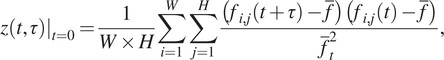
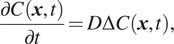
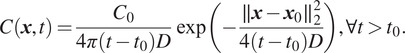
The dynamics and fusion of vesicles during the last steps of exocytosis are not well established yet in cell biology. An open issue is the characterization of the diffusion process at the plasma membrane. Total internal reflection fluorescence microscopy (TIRFM) has been successfully used to analyze the coordination of proteins involved in this mechanism. It enables to capture dynamics of proteins with high frame rate and reasonable signal-to-noise values. Nevertheless, methodological approaches that can analyze and estimate diffusion in local small areas at the scale of a single diffusing spot within cells, are still lacking. To address this issue, we propose a novel correlation-based method for local diffusion estimation. As a starting point, we consider Fick's second law of diffusion that relates the diffusive flux to the gradient of the concentration. Then, we derive an explicit parametric model which is further fitted to time-correlation signals computed from regions of interest (ROI) containing individual spots. Our modeling and Bayesian estimation framework are well appropriate to represent isolated diffusion events and are robust to noise, ROI sizes, and localization of spots in ROIs. The performance of BayesTICS is shown on both synthetic and real TIRFM images depicting Transferrin Receptor proteins.


 分享
分享

 求助内容:
求助内容: 应助结果提醒方式:
 扫码关注我们
