Transcriptomics and metabolomics study in mouse kidney of the molecular mechanism underlying energy metabolism response to hypoxic stress in highland areas.
{"title":"Transcriptomics and metabolomics study in mouse kidney of the molecular mechanism underlying energy metabolism response to hypoxic stress in highland areas.","authors":"Yujie Gao, Qifu Long, Hui Yang, Ying Hu, Yuzhen Xu, Chaoqun Tang, Cunlin Gu, Sheng Yong","doi":"10.3892/etm.2023.12232","DOIUrl":null,"url":null,"abstract":"<p><p>Exposure to hypoxia disrupts energy metabolism and induces inflammation. However, the pathways and mechanisms underlying energy metabolism disorders caused by hypoxic conditions remain unclear. In the present study, a hypoxic animal model was created and transcriptomic and non-targeted metabolomics techniques were applied to further investigate the pathways and mechanisms of hypoxia exposure that disrupt energy metabolism. Transcriptome results showed that 3,007 genes were significantly differentially expressed under hypoxic exposure, and Gene Ontology annotation analysis and Kyoto Encyclopaedia of Genes and Genomes (KEGG) enrichment analysis showed that the differentially expressed genes (DEGs) were mainly involved in energy metabolism and were significantly enriched in the tricarboxylic acid (TCA) cycle and oxidative phosphorylation (OXPHOS) pathway. The DEGs <i>IDH3A</i>, <i>SUCLA2</i>, and <i>MDH2</i> in the TCA cycle and the DEGs <i>NDUFA3</i>, <i>NDUFS7</i>, <i>UQCRC1</i>, <i>CYC1</i> and <i>UQCRFS1</i> in the OXPHOS pathway were validated using mRNA and protein expression, and the results showed downregulation. The results of non-targeted metabolomics showed that 365 significant differential metabolites were identified under plateau hypoxia stress. KEGG enrichment analysis showed that the differential metabolites were mainly enriched in metabolic processes, such as energy, nucleotide and amino acid metabolism. Hypoxia exposure disrupted the TCA cycle and reduced the synthesis of amino acids and nucleotides by decreasing the concentration of cis-aconitate, α-ketoglutarate, NADH, NADPH and that of most amino acids, purines, and pyrimidines. Bioinformatics analysis was used to identify inflammatory genes related to hypoxia exposure and some of them were selected for verification. It was shown that the mRNA and protein expression levels of <i>IL1B</i>, <i>IL12B</i>, <i>S100A8</i> and <i>S100A9</i> in kidney tissues were upregulated under hypoxic exposure. The results suggest that hypoxia exposure inhibits the TCA cycle and the OXPHOS signalling pathway by inhibiting <i>IDH3A</i>, <i>SUCLA2</i>, <i>MDH2</i>, <i>NDUFFA3</i>, <i>NDUFS7</i>, <i>UQCRC1</i>, <i>CYC1</i> and <i>UQCRFS1</i>, thereby suppressing energy metabolism, inducing amino acid and nucleotide deficiency and promoting inflammation, ultimately leading to kidney damage.</p>","PeriodicalId":94002,"journal":{"name":"Experimental and therapeutic medicine","volume":"26 5","pages":"533"},"PeriodicalIF":2.3000,"publicationDate":"2023-09-28","publicationTypes":"Journal Article","fieldsOfStudy":null,"isOpenAccess":false,"openAccessPdf":"https://ftp.ncbi.nlm.nih.gov/pub/pmc/oa_pdf/c3/6e/etm-26-05-12232.PMC10587886.pdf","citationCount":"0","resultStr":null,"platform":"Semanticscholar","paperid":null,"PeriodicalName":"Experimental and therapeutic medicine","FirstCategoryId":"1085","ListUrlMain":"https://doi.org/10.3892/etm.2023.12232","RegionNum":0,"RegionCategory":null,"ArticlePicture":[],"TitleCN":null,"AbstractTextCN":null,"PMCID":null,"EPubDate":"2023/11/1 0:00:00","PubModel":"eCollection","JCR":"","JCRName":"","Score":null,"Total":0}
引用次数: 0
Abstract
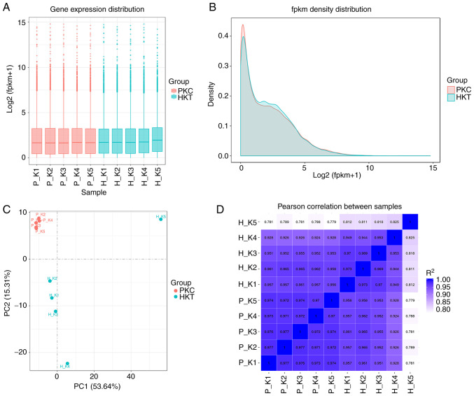
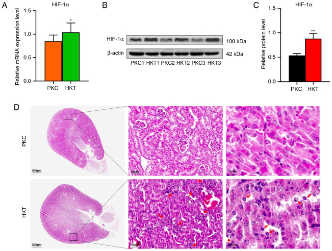
Exposure to hypoxia disrupts energy metabolism and induces inflammation. However, the pathways and mechanisms underlying energy metabolism disorders caused by hypoxic conditions remain unclear. In the present study, a hypoxic animal model was created and transcriptomic and non-targeted metabolomics techniques were applied to further investigate the pathways and mechanisms of hypoxia exposure that disrupt energy metabolism. Transcriptome results showed that 3,007 genes were significantly differentially expressed under hypoxic exposure, and Gene Ontology annotation analysis and Kyoto Encyclopaedia of Genes and Genomes (KEGG) enrichment analysis showed that the differentially expressed genes (DEGs) were mainly involved in energy metabolism and were significantly enriched in the tricarboxylic acid (TCA) cycle and oxidative phosphorylation (OXPHOS) pathway. The DEGs IDH3A, SUCLA2, and MDH2 in the TCA cycle and the DEGs NDUFA3, NDUFS7, UQCRC1, CYC1 and UQCRFS1 in the OXPHOS pathway were validated using mRNA and protein expression, and the results showed downregulation. The results of non-targeted metabolomics showed that 365 significant differential metabolites were identified under plateau hypoxia stress. KEGG enrichment analysis showed that the differential metabolites were mainly enriched in metabolic processes, such as energy, nucleotide and amino acid metabolism. Hypoxia exposure disrupted the TCA cycle and reduced the synthesis of amino acids and nucleotides by decreasing the concentration of cis-aconitate, α-ketoglutarate, NADH, NADPH and that of most amino acids, purines, and pyrimidines. Bioinformatics analysis was used to identify inflammatory genes related to hypoxia exposure and some of them were selected for verification. It was shown that the mRNA and protein expression levels of IL1B, IL12B, S100A8 and S100A9 in kidney tissues were upregulated under hypoxic exposure. The results suggest that hypoxia exposure inhibits the TCA cycle and the OXPHOS signalling pathway by inhibiting IDH3A, SUCLA2, MDH2, NDUFFA3, NDUFS7, UQCRC1, CYC1 and UQCRFS1, thereby suppressing energy metabolism, inducing amino acid and nucleotide deficiency and promoting inflammation, ultimately leading to kidney damage.


 分享
分享

 求助内容:
求助内容: 应助结果提醒方式:
 扫码关注我们
