Identification of Differences in Body Composition Measures Using 3D-Derived Artificial Intelligence from Multiple CT Scans across the L3 Vertebra Compared to a Single Mid-Point L3 CT Scan.
Ke Cao, Josephine Yeung, Yasser Arafat, Matthew Y K Wei, Justin M C Yeung, Paul N Baird
{"title":"Identification of Differences in Body Composition Measures Using 3D-Derived Artificial Intelligence from Multiple CT Scans across the L3 Vertebra Compared to a Single Mid-Point L3 CT Scan.","authors":"Ke Cao, Josephine Yeung, Yasser Arafat, Matthew Y K Wei, Justin M C Yeung, Paul N Baird","doi":"10.1155/2023/1047314","DOIUrl":null,"url":null,"abstract":"<p><strong>Purpose: </strong>Body composition analysis in colorectal cancer (CRC) typically utilises a single 2D-abdominal axial CT slice taken at the mid-L3 level. The use of artificial intelligence (AI) allows for analysis of the entire L3 vertebra (non-mid-L3 and mid-L3). The goal of this study was to determine if the use of an AI approach offered any additional information on capturing body composition measures.</p><p><strong>Methods: </strong>A total of 2203 axial CT slices of the entire L3 level (4-46 slices were available per patient) were retrospectively collected from 203 CRC patients treated at Western Health, Melbourne (97 males; 47.8%). A pretrained artificial intelligence (AI) model was used to segment muscle, visceral adipose tissue (VAT), and subcutaneous adipose tissue (SAT) on these slices. The difference in body composition measures between mid-L3 and non-mid-L3 scans was compared for each patient, and for males and females separately.</p><p><strong>Results: </strong>Body composition measures derived from non-mid-L3 scans exhibited a median range of 0.85% to 6.28% (average percent difference) when compared to the use of a single mid-L3 scan. Significant variation in the VAT surface area (<i>p</i> = 0.02) was observed in females compared to males, whereas male patients exhibited a greater variation in SAT surface area (<i>p</i> < 0.001) and radiodensity (<i>p</i> = 0.007).</p><p><strong>Conclusion: </strong>Significant differences in various body composition measures were observed when comparing non-mid-L3 slices to only the mid-L3 slice. Researchers should be aware that considering only the use of a single midpoint L3 CT scan slice will impact the estimate of body composition measurements.</p>","PeriodicalId":51864,"journal":{"name":"Radiology Research and Practice","volume":"2023 ","pages":"1047314"},"PeriodicalIF":1.5000,"publicationDate":"2023-10-17","publicationTypes":"Journal Article","fieldsOfStudy":null,"isOpenAccess":false,"openAccessPdf":"https://www.ncbi.nlm.nih.gov/pmc/articles/PMC10597731/pdf/","citationCount":"0","resultStr":null,"platform":"Semanticscholar","paperid":null,"PeriodicalName":"Radiology Research and Practice","FirstCategoryId":"1085","ListUrlMain":"https://doi.org/10.1155/2023/1047314","RegionNum":0,"RegionCategory":null,"ArticlePicture":[],"TitleCN":null,"AbstractTextCN":null,"PMCID":null,"EPubDate":"2023/1/1 0:00:00","PubModel":"eCollection","JCR":"Q2","JCRName":"RADIOLOGY, NUCLEAR MEDICINE & MEDICAL IMAGING","Score":null,"Total":0}
引用次数: 0
Abstract
Purpose: Body composition analysis in colorectal cancer (CRC) typically utilises a single 2D-abdominal axial CT slice taken at the mid-L3 level. The use of artificial intelligence (AI) allows for analysis of the entire L3 vertebra (non-mid-L3 and mid-L3). The goal of this study was to determine if the use of an AI approach offered any additional information on capturing body composition measures.
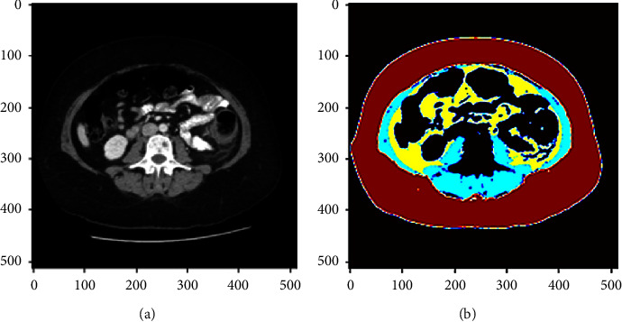
Methods: A total of 2203 axial CT slices of the entire L3 level (4-46 slices were available per patient) were retrospectively collected from 203 CRC patients treated at Western Health, Melbourne (97 males; 47.8%). A pretrained artificial intelligence (AI) model was used to segment muscle, visceral adipose tissue (VAT), and subcutaneous adipose tissue (SAT) on these slices. The difference in body composition measures between mid-L3 and non-mid-L3 scans was compared for each patient, and for males and females separately.
Results: Body composition measures derived from non-mid-L3 scans exhibited a median range of 0.85% to 6.28% (average percent difference) when compared to the use of a single mid-L3 scan. Significant variation in the VAT surface area (p = 0.02) was observed in females compared to males, whereas male patients exhibited a greater variation in SAT surface area (p < 0.001) and radiodensity (p = 0.007).
Conclusion: Significant differences in various body composition measures were observed when comparing non-mid-L3 slices to only the mid-L3 slice. Researchers should be aware that considering only the use of a single midpoint L3 CT scan slice will impact the estimate of body composition measurements.
期刊介绍:
Radiology Research and Practice is a peer-reviewed, Open Access journal that publishes articles on all areas of medical imaging. The journal promotes evidence-based radiology practice though the publication of original research, reviews, and clinical studies for a multidisciplinary audience. Radiology Research and Practice is archived in Portico, which provides permanent archiving for electronic scholarly journals, as well as via the LOCKSS initiative. It operates a fully open access publishing model which allows open global access to its published content. This model is supported through Article Processing Charges. For more information on Article Processing charges in gen


 分享
分享
 求助内容:
求助内容: 应助结果提醒方式:
 扫码关注我们
